. 2014 Oct 28:5:414.
doi: 10.3389/fphys.2014.00414. eCollection 2014.

Oscillatory behavior of ventricular action potential duration in heart failure patients at respiratory rate and low frequency

Affiliations

Oscillatory behavior of ventricular action potential duration in heart failure patients at respiratory rate and low frequency

Ben Hanson et al. Front Physiol. .

Abstract

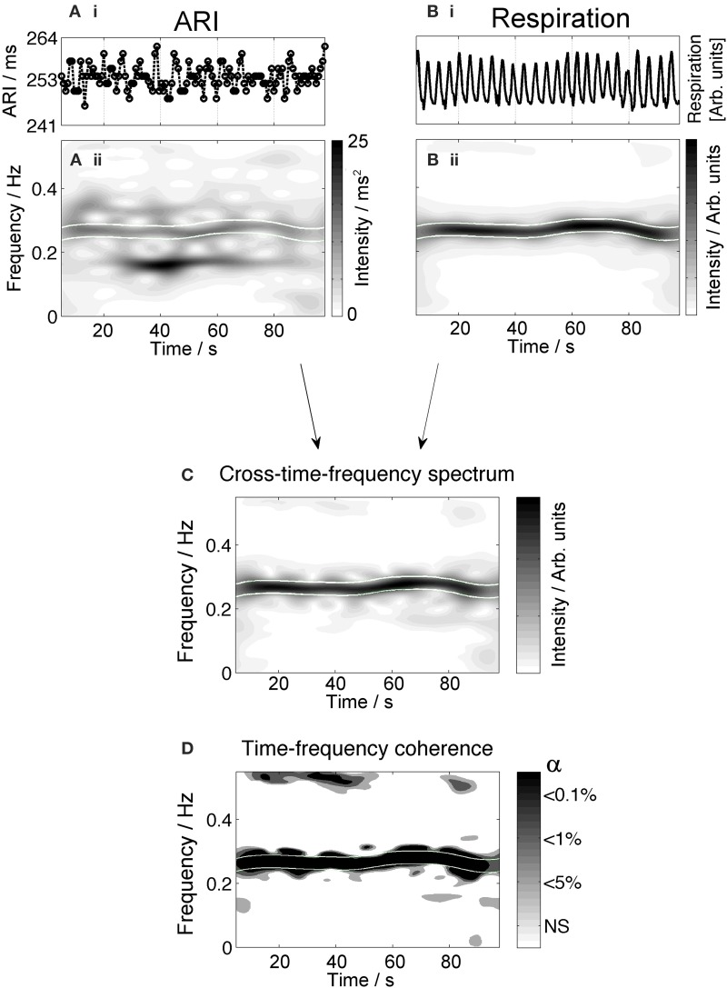
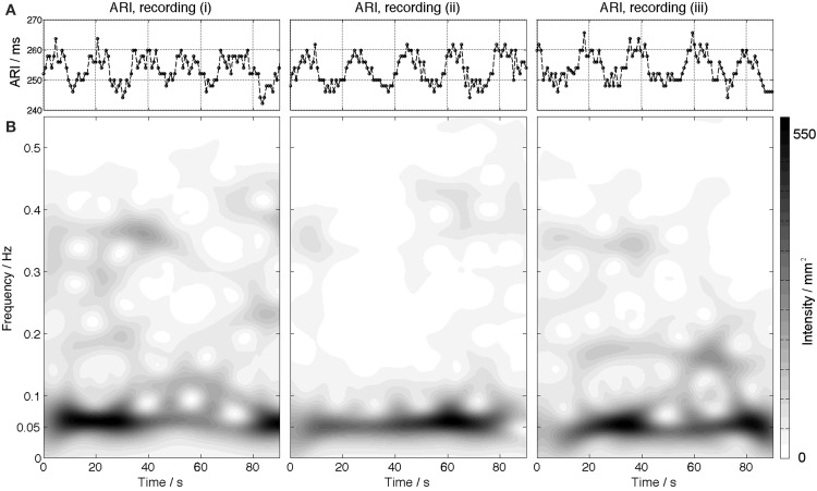
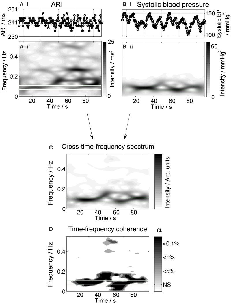
Oscillations of arterial pressure occur spontaneously at a frequency of approximately 0.1 Hz coupled with synchronous oscillations of sympathetic nerve activity ("Mayer waves"). This study investigated the extent to which corresponding oscillations may occur in ventricular action potential duration (APD). Fourteen ambulatory (outpatient) heart failure patients with biventricular pacing devices were studied while seated upright watching movie clips to maintain arousal. Activation recovery intervals (ARI) as a measure of ventricular APD were obtained from unipolar electrograms recorded from the LV epicardial pacing lead during steady state RV pacing from the device. Arterial blood pressure was measured non-invasively (Finapress) and respiration monitored. Oscillations were quantified using time frequency and coherence analysis. Oscillatory behavior of ARI at the respiratory frequency was observed in all subjects. The magnitude of the ARI variation ranged from 2.2 to 6.9 ms (mean 5.0 ms). Coherence analysis showed a correlation with respiratory oscillation for an average of 43% of the recording time at a significance level of p < 0.05. Oscillations in systolic blood pressure in the Mayer wave frequency range were observed in all subjects for whom blood pressure was recorded (n = 13). ARI oscillation in the Mayer wave frequency range was observed in 6/13 subjects (46%) over a range of 2.9 to 9.2 ms. Coherence with Mayer waves at the p < 0.05 significance level was present for an average of 29% of the recording time. In ambulatory patients with heart failure during enhanced mental arousal, left ventricular epicardial APD (ARI) oscillated at the respiratory frequency (approximately 0.25 Hz). In 6 patients (46%) APD oscillated at the slower Mayer wave frequency (approximately 0.1 Hz). These findings may be important in understanding sympathetic activity-related arrhythmogenesis.

Keywords: ARI; Mayer wave; action potential duration; oscillation; respiration.


Figures

Figure 1
Figure 1
Illustration of time-frequency analysis of the respiratory signal and the definition of the respiratory frequency band. The upper panel (A) shows the time series of the respiratory signal. The corresponding time-frequency spectrum is presented in the middle panel (B). The intensity of a specific frequency is given by the grayscale (white: low intensity, black: high intensity). An enhanced frequency band is clearly visible at the respiratory frequency (approximately 0.28 Hz in this example). The frequency spectrum (C) provides a cross-section of the time-frequency plot at 60 s, shown by a dashed line in (B). The respiratory frequency band is defined by taking the maximum amplitude frequency ± the spectral resolution.
Figure 2
Figure 2
Example plot showing oscillations in the ARI signal at the respiratory frequency. The top graphs (Ai,Bi) show the time series of ARI and respiration. The corresponding time-frequency spectra are plotted below, (Aii,Bii). The high intensity band in the time-frequency spectrum of the respiratory signal (Bii) represents the frequency of the respiratory signal (approximately 0.25 Hz). High intensity is also seen in the ARI time-frequency plot (Aii) in this frequency band. Oscillation is also present at other frequencies and times across the spectrum. The cross time- frequency spectrum (C) shows that the ARI and respiratory signal are correlated at the respiratory frequency and the other variations in each signal are not correlated. The results of coherence analysis in the lower panel (D) show the coherence at the respiratory frequency is significant at p < 0.001, indicating that both signals are coupled at this frequency. NS = not significant.
Figure 3
Figure 3
An example is shown for one subject illustrating ARI time series (A) oscillating with a peak-to-peak amplitude of 10–15 ms. The lower panels (B) show the corresponding time-frequency spectra. The spectra show an increased intensity at a frequency of 0.05 Hz.
Figure 4
Figure 4
Example measurements from one subject showing oscillatory behavior of ARI at the Mayer frequency. The upper panels (Ai,Bi) show the time series of ARI and blood pressure, respectively. Blood pressure shows prominent oscillation with a 10-s period; ARI shows oscillation at this rate as well as variation at higher frequencies. The corresponding time-frequency spectra, (Aii,Bii), show high-intensity bands highlighting the presence of waves at a Mayer wave frequency (0.1 Hz). The cross time-frequency spectrum (C) demonstrates that the ARI and blood pressure are correlated at the Mayer frequency and not at other frequencies of oscillation. The results of coherence analysis in the lower panel (D) show the coherence at the respiratory frequency is significant at p < 0.001, indicating that both signals are coupled at this frequency over most of the recording.

References

    1. Anrep G. V., Pascual W., Rossler R. (1936). Respiratory variations of the heart rate. I.-The reflex mechanism of the respiratory arrhythmia. Proc. R. Soc. Lond. B 119B, 191–217. 10.1098/rspb.1936.0005 - DOI
    1. Cevese A., Gulli G., Polati E., Gottin L., Grasso R. (2001). Baroreflex and oscillation of heart period at 0.1 Hz studied by alpha-blockade and cross-spectral analysis in healthy humans. J. Physiol. (Lond.) 531(Pt 1), 235–244. 10.1111/j.1469-7793.2001.0235j.x - DOI - PMC - PubMed
    1. Child N., Hanson B., Bishop M., Rinaldi C. A., Bostock J., Western D., et al. . (2014). Effect of mental challenge induced by movie clips on action potential duration in normal human subjects independent of heart rate. Circ. Arrhythm. Electrophysiol. 15, 2014. 10.1161/CIRCEP.113.000909 - DOI - PMC - PubMed
    1. Cohen M. A., Taylor J. A. (2002). Short-term cardiovascular oscillations in man: measuring and modelling the physiologies. J. Physiol. (Lond.) 542(Pt 3), 669–683. 10.1113/jphysiol.2002.017483 - DOI - PMC - PubMed
    1. Coronel R., de Bakker J. M., Wilms-Schopman F. J., Opthof T., Linnenbank A. C., Belterman C. N., et al. . (2006). Monophasic action potentials and activation recovery intervals as measures of ventricular action potential duration: experimental evidence to resolve some controversies. Heart Rhythm 3, 1043–1050. 10.1016/j.hrthm.2006.05.027 - DOI - PubMed

LinkOut - more resources