

Matching the decay half-life with the biological half-life: ImmunoPET imaging with (44)Sc-labeled cetuximab Fab fragment
- PMID: 25389697
- PMCID: PMC4275156
- DOI: 10.1021/bc500415x
Matching the decay half-life with the biological half-life: ImmunoPET imaging with (44)Sc-labeled cetuximab Fab fragment
Abstract
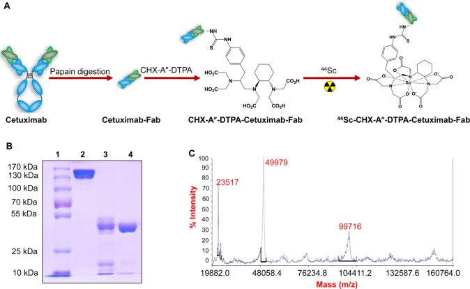
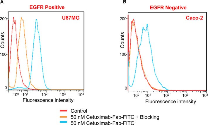
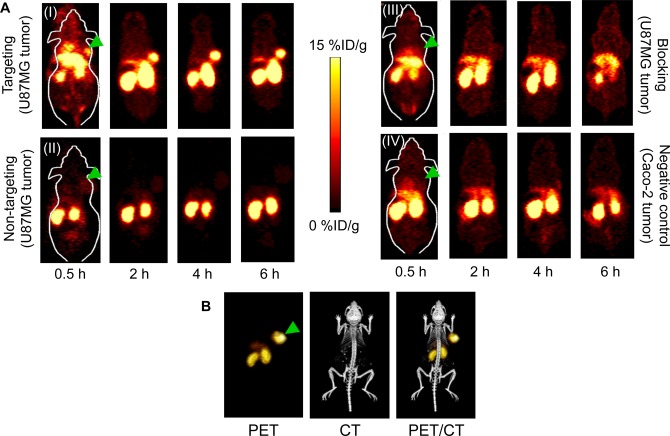
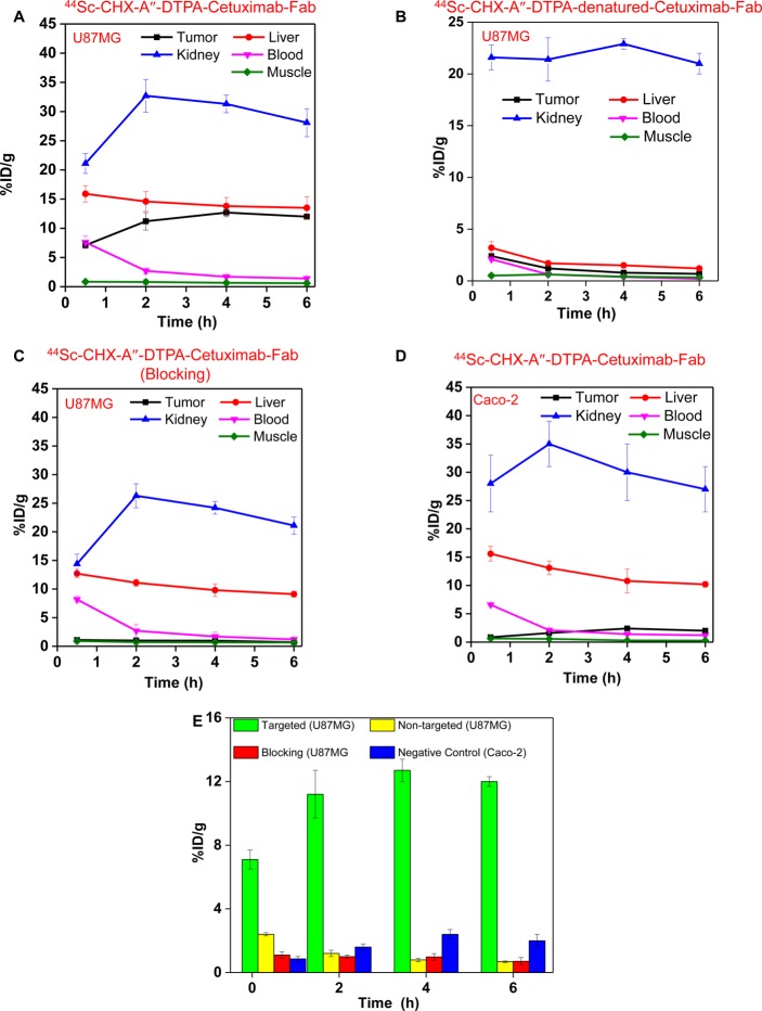
Scandium-44 (t1/2 = 3.9 h) is a relatively new radioisotope of potential interest for use in clinical positron emission tomography (PET). Herein, we report, for the first time, the room-temperature radiolabeling of proteins with (44)Sc for in vivo PET imaging. For this purpose, the Fab fragment of Cetuximab, a monoclonal antibody that binds with high affinity to epidermal growth factor receptor (EGFR), was generated and conjugated with N-[(R)-2-amino-3-(para-isothiocyanato-phenyl)propyl]-trans-(S,S)-cyclohexane-1,2-diamine-N,N,N',N″,N″-pentaacetic acid (CHX-A″-DTPA). The high purity of Cetuximab-Fab was confirmed by SDS-PAGE and mass spectrometry. The potential of the bioconjugate for PET imaging of EGFR expression in human glioblastoma (U87MG) tumor-bearing mice was investigated after (44)Sc labeling. PET imaging revealed rapid tumor uptake (maximum uptake of ∼12% ID/g at 4 h postinjection) of (44)Sc-CHX-A″-DTPA-Cetuximab-Fab with excellent tumor-to-background ratio, which might allow for same day PET imaging in future clinical studies. Immunofluorescence staining was conducted to correlate tracer uptake in the tumor and normal tissues with EGFR expression. This successful strategy for immunoPET imaging of EGFR expression using (44)Sc-CHX-A″-DTPA-Cetuximab-Fab can make clinically translatable advances to select the right population of patients for EGFR-targeted therapy and also to monitor the therapeutic efficacy of anti-EGFR treatments.
Figures

References
-
- Roesch F. (2012) Scandium-44: benefits of a long-lived PET radionuclide available from the 44Ti/44Sc generator system. Curr. Radiopharm. 5, 187–201. - PubMed
-
- Cutler C. S.; Hennkens H. M.; Sisay N.; Huclier-Markai S.; Jurisson S. S. (2013) Radiometals for combined imaging and therapy. Chem. Rev. 113, 858–83. - PubMed
-
- Koumarianou E.; Loktionova N. S.; Fellner M.; Roesch F.; Thews O.; Pawlak D.; Archimandritis S. C.; Mikolajczak R. (2012) 44Sc-DOTA-BN[2–14]NH2 in comparison to 68Ga-DOTA-BN[2–14]NH2 in pre-clinical investigation. Is 44Sc a potential radionuclide for PET?. Appl. Radiat. Isot. 70, 2669–76. - PubMed
-
- Eigner S.; Vera D. R.; Fellner M.; Loktionova N. S.; Piel M.; Lebeda O.; Rosch F.; Ross T. L.; Henke K. E. (2013) Imaging of protein synthesis: in vitro and in vivo evaluation of 44Sc-DOTA-puromycin. Mol. Imaging Biol. 15, 79–86. - PubMed
Publication types
MeSH terms
Substances
Grants and funding
LinkOut - more resources
Full Text Sources
Other Literature Sources
Research Materials
Miscellaneous
