Review
. 2014 Nov 10;6(11):4984-5017.
doi: 10.3390/nu6114984.

Vitamin a deficiency and alterations in the extracellular matrix

Affiliations
Review

Vitamin a deficiency and alterations in the extracellular matrix

Teresa Barber et al. Nutrients. .

Abstract

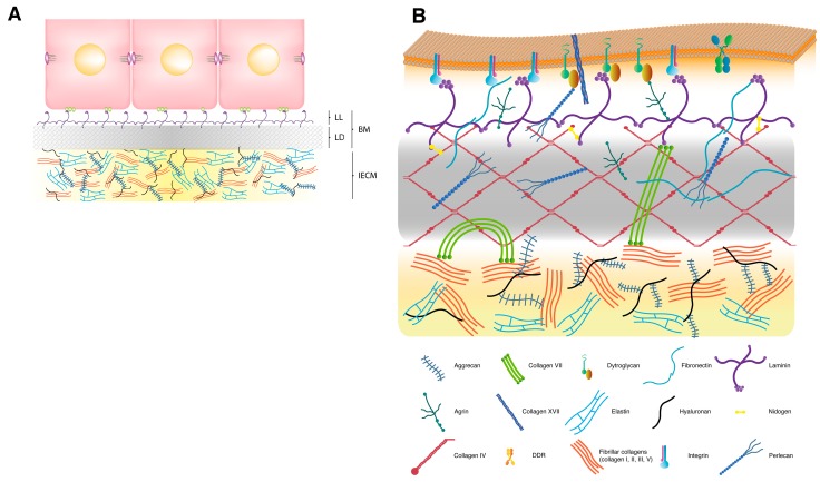
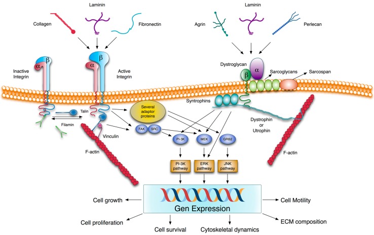
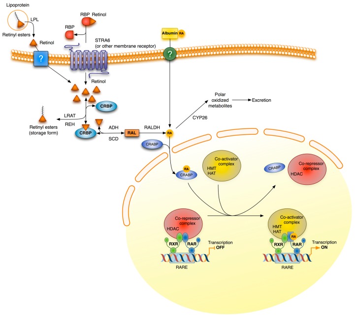
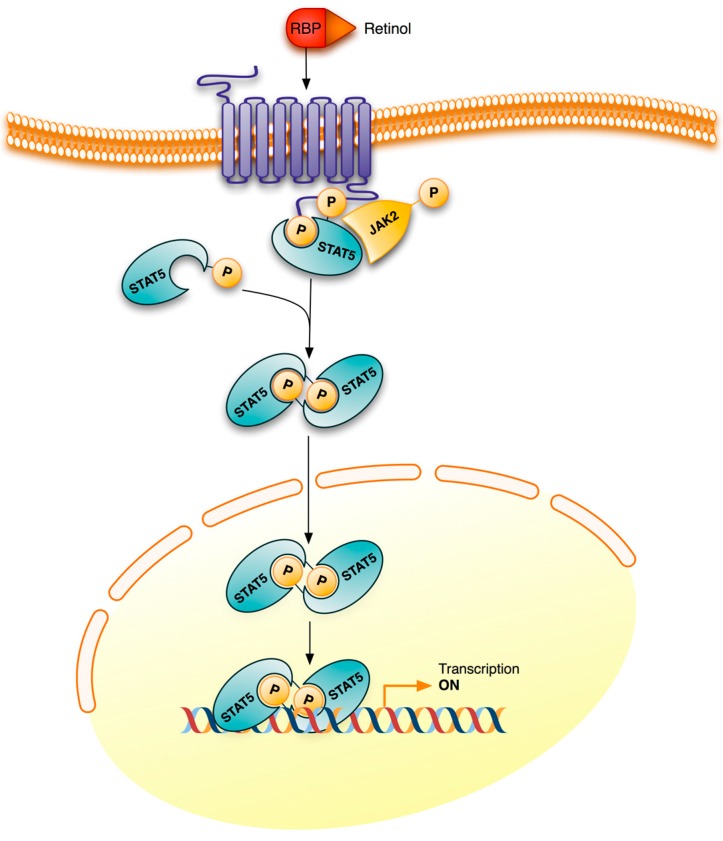
Vitamin A or retinol which is the natural precursor of several biologically active metabolites can be considered the most multifunctional vitamin in mammals. Its deficiency is currently, along with protein malnutrition, the most serious and common nutritional disorder worldwide. It is necessary for normal embryonic development and postnatal tissue homeostasis, and exerts important effects on cell proliferation, differentiation and apoptosis. These actions are produced mainly by regulating the expression of a variety of proteins through transcriptional and non-transcriptional mechanisms. Extracellular matrix proteins are among those whose synthesis is known to be modulated by vitamin A. Retinoic acid, the main biologically active form of vitamin A, influences the expression of collagens, laminins, entactin, fibronectin, elastin and proteoglycans, which are the major components of the extracellular matrix. Consequently, the structure and macromolecular composition of this extracellular compartment is profoundly altered as a result of vitamin A deficiency. As cell behavior, differentiation and apoptosis, and tissue mechanics are influenced by the extracellular matrix, its modifications potentially compromise organ function and may lead to disease. This review focuses on the effects of lack of vitamin A in the extracellular matrix of several organs and discusses possible molecular mechanisms and pathologic implications.


Figures

Figure 1
Figure 1
Schematic model of the extracellular matrix and the basement membrane. (A) Extracellular matrix (ECM). In many tissues, such as glandular, stratified and certain complex epithelia, the ECM is formed by a basement membrane (BM), which is connected to the epithelial cells, and an interstitial extracellular matrix (IECM), which constitutes most ECM space. Cells adopt basal-apical polarity and interact with BM components through specific cell surface receptors located on their basal side. As in transmission electron microscopy after conventional fixation, the BM is composed of two layers: (1) the lamina lucida (LL), an electron-lucent layer that lies immediately adjacent to the cell; and (2) the lamina densa (LD), an electron-dense layer that comes into contact with the IECM. The lamina lucida is not detectable in electron microscopy specimens fixed by the milder method of freeze substitution. (B) Basement membrane. The basic structure of a BM is formed by two independent networks of laminin and collagen IV, which are non-covalently interconnected by nidogen and perlecan. The self-assembled collagen IV network is recruited together with other BM components, including nidogen, perlecan and agrin, to the nascent laminin scaffold. Agrin and perlecan provide additional connections between the BM and the cell surface. Native collagens also have the possibility of contacting cells through plasma membrane receptors, such as DDRs (not shown in the figure). Several molecules, such as collagens VII, XV and XVIII, are known to act as linkers between the BM and the adjacent IECM. For clarity purposes, only collagen VII aggregates, which may interact with fibronectin, laminin, collagen I and collagen IV, and form the anchoring fibrils, are included in the figure.
Figure 2
Figure 2
Integrin- and dystroglycan-mediated ECM signaling. Several ECM components are able to interact with integrins and dystroglycans. Unlike dystroglycan, integrins are present on the cell surface as inactive and active forms. The conformational switch from low to high ligand affinity may be regulated by different ways. These include β-subunit cytoplasmic tail phosphorylation and competitive binding to this β-subunit tail between activators and inhibitors, such as cytoskeletal proteins talin and filamin, respectively. Ligand-integrin binding induces the recruitment of several adaptor proteins and the phosphorylation of some protein kinases such as focal adhesion kinase (FAK) which trigger different signaling pathways. In addition, integrins are linked to actin filaments through proteins, such as talin, filamin or actinin. Similarly, the interaction of α-dystroglycan with the LG domain containing ligands transduces extracellular information via β-dystroglycan to generate intracellular signals, which are mediated by signaling pathways, also activated by integrins. In fact, there is much evidence to suggest a cross-talk between both receptors. The signaling pathways induced by integrins and dystroglycans modulate gene expression patterns and impact cell behavior. The molecular organization of both receptors with their associated proteins provides a physical connection between the ECM and the cytoskeleton, and a way to transmit mechanical cues between them. ERK, extracellular signal-regulated kinase; FAK, focal adhesion kinase; GRB2, growth factor receptor-bound protein 2; JNK, c-Jun N-terminal kinase; MEK, MAPK kinase; PI-3K, phosphatidylinositol 3-kinase; Src, protein tyrosine kinase encoded by the c-src gene.
Figure 3
Figure 3
General scheme of retinoic acid signaling and metabolism. Retinoids enter cells via several possible routes. In extrahepatic tissues, RBP-bound retinol enters cells through the STRA6 receptor or any other related receptor; and free retinol, derived from lipoproteins by the action of lipoprotein lipase, and albumin-bound RA, which is present in small amounts in plasma, may enter by passive diffusion and/or by any uncharacterized receptor. Hepatocytes and, to a lesser extent, also extrahepatic cells, obtain retinyl esters by lipoprotein uptake. Inside cells, retinol is esterified and stored as retinyl esters or is metabolized to retinoic acid (RA) by two sequential oxidations. It is thought that intracellular retinoid-binding proteins, such as cellular retinol-binding proteins (CRBP) and cellular retinoic acid-binding proteins (CRABP), participate in the coordination of these processes. RA is degraded to more polar, less bio-active metabolites by enzymes of the CYP26 family. CRABP-bound RA is translocated to the nucleus where it binds to the retinoic acid receptor (RAR) and initiates gene transcription. This effect is produced even in the absence of an RXR agonist, however the binding of agonists to both receptor partners improves transcription efficiency. ADH, alcohol dehydrogenases; CRBP, cellular retinol-binding protein; CRABP, cellular retinoic acid binding protein; CYP26, family 26 of cytochrome P450 enzymes; HAT, histone acetyltransferase; HDAC, histone deacetylase; HMT, histone methyltransferase; LPL, lipoprotein lipase; LRAT, lecithin retinol acyltransferase; RALDH, retinaldehyde dehydrogenase; RAR, retinoic acid receptor; RARE, retinoic acid response element; RBP, retinol binding protein; REH, retinyl ester hydrolases; RXR, retinoid X receptor; SDR, short-chain dehydrogenase/reductases.
Figure 4
Figure 4
Retinol signaling. STRA6 is not only a plasma membrane transporter for retinol but also a signaling receptor. Binding of the retinol-RBP complex to the extracellular part of STRA6 triggers the phosphorylation of its cytosolic domain. The phosphorylated domain recruits and activates JAK2 which, in turn, phosphorylates STAT5, also bound to the phosphorylated receptor. Phosphorylated STAT5 dimers initiate transcription of their target genes. The letter P inside a circle denotes a phosphoryl group. JAK2, Janus kinase 2; RBP, retinol binding protein; STAT5, signal transducer and activator of transcription 5; STRA6, stimulated by retinoic acid 6.

Similar articles

Cited by

References

    1. McLaren D.S., Kraemer K. Vitamin A in nature. World Rev. Nutr. Diet. 2012;103:7–17. - PubMed
    1. De Luca L.M. Retinoids and their receptors in differentiation, embryogenesis and neoplasia. FASEB J. 1991;5:2924–2933. - PubMed
    1. Duester G. Retinoic acid synthesis and signaling during early organogenesis. Cell. 2008;134:921–931. doi: 10.1016/j.cell.2008.09.002. - DOI - PMC - PubMed
    1. Mark M., Ghyselinck N.B., Chambon P. Function of retinoic acid receptors during embryonic development. Nucl. Recept. Signal. 2009;7:e002. - PMC - PubMed
    1. Ross A.C. Vitamin A and retinoic acid in T cell-related immunity. Am. J. Clin. Nutr. 2012;96:1166S–1172S. doi: 10.3945/ajcn.112.034637. - DOI - PMC - PubMed

MeSH terms