. 2015 Feb 15;191(4):417-26.
doi: 10.1164/rccm.201406-1162OC.

Extensive phenotyping of individuals at risk for familial interstitial pneumonia reveals clues to the pathogenesis of interstitial lung disease

Affiliations

Extensive phenotyping of individuals at risk for familial interstitial pneumonia reveals clues to the pathogenesis of interstitial lung disease

Jonathan A Kropski et al. Am J Respir Crit Care Med. .

Abstract

Rationale: Asymptomatic relatives of patients with familial interstitial pneumonia (FIP), the inherited form of idiopathic interstitial pneumonia, carry increased risk for developing interstitial lung disease.

Objectives: Studying these at-risk individuals provides a unique opportunity to investigate early stages of FIP pathogenesis and develop predictive models of disease onset.

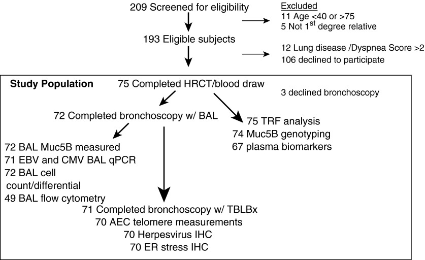
Methods: Seventy-five asymptomatic first-degree relatives of FIP patients (mean age, 50.8 yr) underwent blood sampling and high-resolution chest computed tomography (HRCT) scanning in an ongoing cohort study; 72 consented to bronchoscopy with bronchoalveolar lavage (BAL) and transbronchial biopsies. Twenty-seven healthy individuals were used as control subjects.

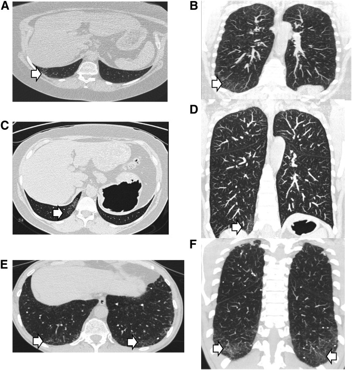
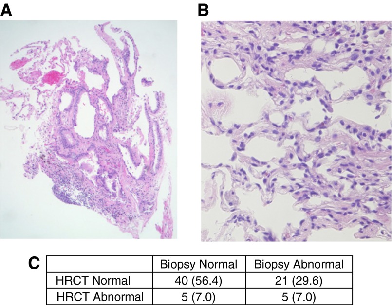
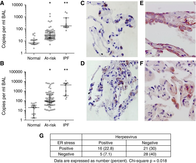
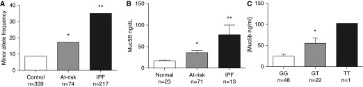
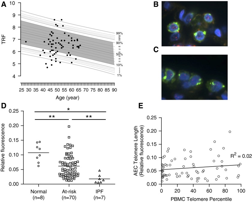
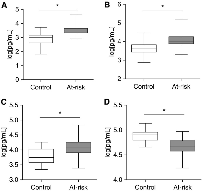
Measurements and main results: Eleven of 75 at-risk subjects (14%) had evidence of interstitial changes by HRCT, whereas 35.2% had abnormalities on transbronchial biopsies. No differences were noted in inflammatory cells in BAL between at-risk individuals and control subjects. At-risk subjects had increased herpesvirus DNA in cell-free BAL and evidence of herpesvirus antigen expression in alveolar epithelial cells (AECs), which correlated with expression of endoplasmic reticulum stress markers in AECs. Peripheral blood mononuclear cell and AEC telomere length were shorter in at-risk individuals than healthy control subjects. The minor allele frequency of the Muc5B rs35705950 promoter polymorphism was increased in at-risk subjects. Levels of several plasma biomarkers differed between at-risk subjects and control subjects, and correlated with abnormal HRCT scans.

Conclusions: Evidence of lung parenchymal remodeling and epithelial dysfunction was identified in asymptomatic individuals at risk for FIP. Together, these findings offer new insights into the early pathogenesis of idiopathic interstitial pneumonia and provide an ongoing opportunity to characterize presymptomatic abnormalities that predict progression to clinical disease.

Keywords: IPF; alveolar epithelial cell; biomarker; bronchoscopy; telomere.


Figures

Figure 1.
Figure 1.
Schematic of enrollment and study population. AEC = alveolar epithelial cells; BAL = bronchoalveolar lavage; CMV = cytomegalovirus; EBV = Epstein-Barr virus; ER = endoplasmic reticulum; HRCT = high-resolution computed tomography; IHC = immunohistochemistry; qPCR = quantitative polymerase chain reaction; TBLBx = transbronchial lung biopsies; TRF = telomere restriction fragment.
Figure 2.
Figure 2.
High-resolution computed tomography abnormalities in at-risk subjects. Axial and coronal images of three different individuals demonstrating representative radiographic abnormalities are shown by arrows. (A and B) Intralobular reticular opacities. (C and D) Irregular intralobular septal thickening. (E and F) Traction bronchiolectasis.
Figure 3.
Figure 3.
Transbronchial biopsy histology from at-risk subjects. Abnormal features identified on transbronchial biopsy specimens include (A) coarse interstitial fibrosis associated with normal tissue, hyperplastic epithelial cells and (B) peribronchiolar metaplasia with perivascular inflammation. (C) Association between high-resolution computed tomography (HRCT) and transbronchial biopsies among 71 subjects. Original magnification (A) 40×, and (B) 200×.
Figure 4.
Figure 4.
Herpesviruses are detectable in lungs from at-risk subjects. DNA was isolated from cell-free bronchoalveolar lavage (BAL) supernatant from normal control subjects, subjects at-risk for familial interstitial pneumonia, and idiopathic pulmonary fibrosis (IPF) control subjects. Copies of Epstein-Barr virus (EBV) (A) and cytomegalovirus (CMV) (B) were quantified by quantitative polymerase chain reaction (bar represents median and interquartile range). EBV (C) and CMV (D) antigens were also detected by immunostaining in type II alveolar epithelial cells in transbronchial biopsy specimens from at-risk subjects. (E) Bip and (F) XBP1 were identified in alveolar epithelial cells in transbronchial biopsies from at-risk subjects. (G) Quantification of transbronchial biopsy specimens from at-risk subjects stained for endoplasmic reticulum (ER) stress markers Bip and XBP1 and herpesvirus antigens from EBV and CMV. *P < 0.001 at-risk versus normal control subjects. **P < 0.001 IPF versus normal control subjects.
Figure 5.
Figure 5.
Muc5B promoter polymorphism rs35705950 is found with increased frequency in at-risk subjects. (A) At-risk subjects along with 339 healthy control subjects and 217 idiopathic pulmonary fibrosis (IPF) patients were genotyped for the Muc5B rs35705950 promoter polymorphism. Minor allele frequency is increased in at-risk subjects compared with control subjects. (B) Muc5B level in bronchoalveolar lavage supernatant was measured by ELISA in normal control subjects, at-risk subjects, and IPF control subjects. (C) Muc5B levels in homozygote major allele carriers (GG), heterozygote minor allele carriers (GT), and homozygote minor allele carriers (TT). *P < 0.05 at-risk versus normal control subjects. **P < 0.05 IPF versus normal control subjects.
Figure 6.
Figure 6.
At-risk subjects have short telomeres in peripheral blood and type II alveolar epithelial cells. (A) DNA was isolated from peripheral blood mononuclear cells (PBMCs) of at-risk subjects and telomere restriction fragment (TRF) was measured by Southern blot. TRF was then plotted against age in comparison with a population of normal control subjects. Telomeres less than the 10th percentile for age were considered abnormally short. (B and C) Telomere length in type II alveolar epithelial cells (AECs) was measured using a dual-fluorescence approach wherein type II AECs were identified by costaining for pro–surfactant protein C, and telomeric DNA was quantified relative to total nuclear DNA (DAPI) by relative fluorescence in (B) control and (C) at-risk subjects. Green = pro–surfactant protein C; blue = DAPI (nuclear DNA); red within nucleus = telomeres. (D) Quantification of telomere fluorescence intensity relative to nuclear DNA in type II AECs. *P < 0.001, **P < 0.005. (E) PBMC telomere length was plotted versus AEC telomere length and a linear regression was performed to determine the relationship between PBMC and AEC telomere length. IPF = idiopathic pulmonary fibrosis.
Figure 7.
Figure 7.
Plasma biomarkers differ between at-risk subjects and healthy control subjects. Plasma levels of (A) matrix metalloproteinase 7, (B) surfactant protein-D, (C) ET-1, and (D) TIMP2 in 67 at-risk and 27 healthy control subjects. Data were log transformed to normalize distribution for analysis. Boxes indicate 25th–75th percentiles; whiskers depict minimum to maximum values. At-risk and control subjects were compared using a mixed-effects model controlling for age and sex. *P < 0.001 is considered significant after Bonferroni correction for multiple comparisons.

Comment in

References

    1. García-Sancho C, Buendía-Roldán I, Fernández-Plata MR, Navarro C, Pérez-Padilla R, Vargas MH, Loyd JE, Selman M. Familial pulmonary fibrosis is the strongest risk factor for idiopathic pulmonary fibrosis. Respir Med. 2011;105:1902–1907. - PubMed
    1. Loyd JE. Pulmonary fibrosis in families. Am J Respir Cell Mol Biol. 2003;29(3) Suppl:S47–S50. - PubMed
    1. Kropski JA, Lawson WE, Young LR, Blackwell TS. Genetic studies provide clues on the pathogenesis of idiopathic pulmonary fibrosis. Dis Model Mech. 2013;6:9–17. - PMC - PubMed
    1. Lawson WE, Loyd JE, Degryse AL. Genetics in pulmonary fibrosis—familial cases provide clues to the pathogenesis of idiopathic pulmonary fibrosis. Am J Med Sci. 2011;341:439–443. - PMC - PubMed
    1. Steele MP, Speer MC, Loyd JE, Brown KK, Herron A, Slifer SH, Burch LH, Wahidi MM, Phillips JA, III, Sporn TA, et al. Clinical and pathologic features of familial interstitial pneumonia. Am J Respir Crit Care Med. 2005;172:1146–1152. - PMC - PubMed

MeSH terms