. 2014 Nov 12;9(11):e111898.
doi: 10.1371/journal.pone.0111898. eCollection 2014.

Alzheimer's therapeutics targeting amyloid beta 1-42 oligomers I: Abeta 42 oligomer binding to specific neuronal receptors is displaced by drug candidates that improve cognitive deficits

Affiliations

Alzheimer's therapeutics targeting amyloid beta 1-42 oligomers I: Abeta 42 oligomer binding to specific neuronal receptors is displaced by drug candidates that improve cognitive deficits

Nicholas J Izzo et al. PLoS One. .

Abstract

Synaptic dysfunction and loss caused by age-dependent accumulation of synaptotoxic beta amyloid (Abeta) 1-42 oligomers is proposed to underlie cognitive decline in Alzheimer's disease (AD). Alterations in membrane trafficking induced by Abeta oligomers mediates reduction in neuronal surface receptor expression that is the basis for inhibition of electrophysiological measures of synaptic plasticity and thus learning and memory. We have utilized phenotypic screens in mature, in vitro cultures of rat brain cells to identify small molecules which block or prevent the binding and effects of Abeta oligomers. Synthetic Abeta oligomers bind saturably to a single site on neuronal synapses and induce deficits in membrane trafficking in neuronal cultures with an EC50 that corresponds to its binding affinity. The therapeutic lead compounds we have found are pharmacological antagonists of Abeta oligomers, reducing the binding of Abeta oligomers to neurons in vitro, preventing spine loss in neurons and preventing and treating oligomer-induced deficits in membrane trafficking. These molecules are highly brain penetrant and prevent and restore cognitive deficits in mouse models of Alzheimer's disease. Counter-screening these compounds against a broad panel of potential CNS targets revealed they are highly potent and specific ligands of the sigma-2/PGRMC1 receptor. Brain concentrations of the compounds corresponding to greater than 80% receptor occupancy at the sigma-2/PGRMC1 receptor restore cognitive function in transgenic hAPP Swe/Ldn mice. These studies demonstrate that synthetic and human-derived Abeta oligomers act as pharmacologically-behaved ligands at neuronal receptors--i.e. they exhibit saturable binding to a target, they exert a functional effect related to their binding and their displacement by small molecule antagonists blocks their functional effect. The first-in-class small molecule receptor antagonists described here restore memory to normal in multiple AD models and sustain improvement long-term, representing a novel mechanism of action for disease-modifying Alzheimer's therapeutics.


Conflict of interest statement

Competing Interests: NI, HW, TW, AV, MW, ZS, JR, KM, CS, CR, RY, PF, GL, GR, MS, HS, SC, are employees or paid consultants of Cognition Therapeutics. HL owns stock or stock options of Cognition Therapeutics. This does not alter the authors' adherence to PLOS ONE policies on sharing data and materials.

Figures

Figure 1
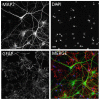
Figure 1. Immunofluorescent labeling of mixed hippocampal/cortical cultures.
A, MAP2 labeled neurons. B, DAPI-labeled nuclei. C, GFAP labeled glia and Nuclei. D, Merged composite of all three images. Based on untreated control wells from 104 experimental plates, the percentage of neurons in the cultures was 26.0±1.1% (Mean ± S.E.M.). Scale bar  = 20 microns.
Figure 2
Figure 2. Characterization of synthetic human Abeta 1–42 oligomers by non-denaturing Western blot, MALDI-TOF.
A, Freshly prepared solutions of synthetic human Abeta 1–42 (lane 1) or 1–40 (lane 3) peptide loaded onto non-denaturing western gels immediately after reconstitution contain large amounts of monomer (arrow; fainter lower molecular weight band represents peptide degradation product) and little higher molecular weight material. In contrast, the same solution of Abeta 1–42 peptide that is allowed to oligomerize for 24 hours (lane 2) contains much larger amounts of higher molecular weight material >50 kDa, and less monomeric protein. The full length of gel lanes are shown from loading well to dye front. Note that oligomers run differently on non-denaturing gels than globular molecular weight protein size standards . B. The presence of significant amounts of monomer in oligomer preparations is also confirmed by MALDI-TOF analysis of the same Abeta 1–42 oligomer preparation that shows both a 4.5 kDa monomer peak and multiple lower abundance peaks corresponding to oligomers of various sizes. MALDI-TOF (detection range 3–100 kDa) of vehicle (media without Abeta) is shown below for comparison (C).
Figure 3
Figure 3. Characterization of human Abeta 1–42 oligomers isolated from patient frozen, unfixed 1 gram brain samples by non-denaturing Western blot, MALDI-TOF and ELISA.
A, Non-denaturing Western blots of immunoprecipitated Alzheimer's patient hippocampal samples demonstrates heterogeneous populations of oligomer assemblies. 6E10 antibody labeling of western blots from four different AD patients (lanes 1–4) detects major bands ≥250 kDa, and multiple discrete bands between 50–75 kDa. In contrast, APP antibody detects a faint band at 125 kDa (lane 5). Significant amounts of monomeric Abeta 1–42 were not observed in any individual. MALDI-TOF analysis of immune-precipitated human brain samples demonstrates heterogeneous populations of oligomer assemblies, both between individual Alzheimer's patients (B, D) and between age-matched histologically normal individuals (C, E). Significant amounts of monomeric Abeta 1–42 were not observed in any individual. Albumin was added to samples as an internal size control (arrow in B–E).
Figure 4
Figure 4. Characterization of Membrane Trafficking Assay.
Membrane trfficking rate is measured by quantifying the number of intracellular vesicles labeled with endocytic cargo dye in mature primary hippocampal cultures (≥21 DIV). Over time, the dye-filled vesicles (A) are trafficked out of cells via exocytosis, and the amount of dye in vesicles decreases as it crystalizes on the cell surface as needle-shaped crystals (B). Abeta oligomer treatment affects the rate of membrane trafficking; 60 minutes following addition of MTT reagent, when vehicle treated neurons (A) still contain labeled vesicles, Abeta oligomer-treated neurons (B) have already exocytosed labeled vesicles. C. Addition of antibody to Abeta (6E10) to cultures prevents Abeta oligomers from affecting trafficking rates. Amount of labeled vesicles is quantified (y-axis in D) as a percentage of vehicle-treated values. D. Concentration-dependent effect of synthetic Abeta oligomer preparation on vesicular labeling relative to vehicle treated cells is blocked by antibody IgG to Abeta. E. Memantine (MEM), MK-801, and scyllo-inositol were run as controls in the membrane trafficking assay.
Figure 5
Figure 5. Relative potency of Abeta preparations in membrane trafficking assay.
Synthetic human Abeta 1–42 oligomer (high concentration), freshly made monomer, synthetic oligomers (low concentration), semi-synthetic oligomers and human Alzheimer's patient derived oligomers were dosed in the membrane trafficking assay. All Abeta preparations alter membrane trafficking rates but with different EC50 concentrations and different exposure times to Bmax, similar to literature reports ( Table 2 ).
Figure 6
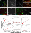
Figure 6. Abeta oligomers bind to a single saturable receptor site on neuronal synaptic puncta.
A, B. Abeta 1–42 oligomers bind to some but not all neurons when added to cultures for 60 minutes (total Abeta concentration  = 440 nM, visualized with 6E10 immunolabeling). A subset of neurons (immunopositive for MAP2, green) exhibit punctate postsynaptic oligomer binding (red) along their neurites; 37%±3% of these puncta colocalize with presynaptic terminals immunolabeled for synaptophysin (C, D). Nuclei (DAPI +, blue) of several non-neuronal cells (glia, MAP2-negative) exhibit Abeta binding to the cell body. B, F, H. Immunolabeling of Abeta, alone, is shown for clarity. E, F. Fresh Abeta 1–42 monomers, added to cells with identical concentrations and conditions, is characterized by very low intensity punctate labeling on neurites and labeling of neuron and glia cell bodies. G, H. 6E10 (monoclonal antibody to Abeta 3–8) added to cultures prior to oligomers blocks binding of Abeta oligomers to neurite puncta but not to cell bodies of neurons or glia. I. Binding isotherms for Abeta oligomers (red closed circles) and fresh Abeta monomer (black open circles; treatment for 60 minutes with 44 nM- 14 µM total Abeta concentration) indicate that oligomer binding to neuronal puncta fits a single-site, saturable model (Kd = 518±41 nM, Table 3 ). Binding of fresh monomer fits a two site model with a high affinity site (412±48 nM) and a second non-saturable binding site. J. Same data as I, expanded to show concentrations used for competition binding studies (440 nM total Abeta concentration); at these low concentrations binding intensity of oligomers to neuronal synaptic puncta is five-fold higher than with monomer. K, L. Binding of synthetic Abeta oligomers and fresh monomer to glial cells with round/symmetric nuclear morphology (closed arrowheads in B, F) is not above background levels (zero Abeta concentration in all graphs). M, N. Binding of synthetic Abeta oligomers and fresh monomer to glia with condensed/asymmetric nuclear morphology(open arrowheads in B, F) fits a two site model with a high affinity site (Kd = 289±150 nM) and a second non-saturating site (Kd>1 M). Scale bars  = 20 µm.
Figure 7
Figure 7. Effect of Abeta control peptides in membrane trafficking assay.
Oligomer preparation of Abeta 1–40 is 200 times less potent than Abeta 1–42 in binding assay (A) and in membrane trafficking (B). Scrambled Abeta 1–42 is not active in the membrane trafficking assay and is not detectable by the antibody used for the Abeta binding assay.
Figure 8
Figure 8. Chemical structures of compounds.
Figure 9
Figure 9. Abeta 1–42 oligomer-induced trafficking deficits are prevented and competitively inhibited by sigma-2/PGRMC1 antagonists.
A, Reduced cargo dye (formazan) labels intracellular vesicles in vehicle-treated cultures, but is trafficked out of the cell more rapidly (B) following addition of synthetic Abeta oligomers (3 µM, total Abeta concentration). 15 µM CT0109 added 1 hr prior to Abeta restores trafficking to vehicle-treated levels (C) without affecting vesicles on its own (D). E–H Dose-response curves showing that CT0109 (E), CT0093 (F), CT01344 (G), and CT01346 (H) restore trafficking deficits whether added before (prevention) or after (treatment) addition of Abeta oligomers. I–L, synthetic Abeta oligomers exhibit a dose-dependent inhibition of membrane trafficking (red) that is right-shifted 5 to10-fold in the presence of increasing concentrations of sigma-2/PGRMC1 antagonists (I, CT0109: EC50 = 0.5 µM to 5.3 µM; J, CT0093: EC50 = 0.81 µM to 12.8 µM; K,CT01344: EC50 = 1.7 µM to 7.1 µM; L, CT01346 EC50 = 5.7 µM to 12.2 µM), consistent with pharmacological competition between compounds and Abeta oligomers. M, N, Abeta oligomers isolated from human postmortem AD patients exhibits a more potent dose-dependent inhibition of membrane trafficking (red), yet the same compounds inhibit the maximum effect of oligomers by 60 to 99% (P<0.025, t-test) making these oligomers less efficacious and therefore less toxic. O, CT0109 and CT0093 (0.2 µM, grey and black filled bars) reverse the trafficking deficit cause by human patient derived Abeta (1.5 pM, red bar) without affecting trafficking on their own (gray and black open bars).
Figure 10
Figure 10. Small molecule therapeutic candidates can prevent or displace Abeta oligomer binding to mature primary hippocampal and cortical cultures (21DIV).
Abeta synthetic oligomers were added to neuronal cultures (0.5 µM total Abeta concentration) for 40 min prior to (A–F) or following (G–L) addition of compounds and bound Abeta was detected by immunofluorescence. A, G Vehicle controls with no Abeta show background fluorescence. C, I, 15 µM CT0109, D, J, CT0093, E, K, CT01344, or F, L, CT01346 show that immuno-fluorescence for punctate binding of Abeta oligomers to neurites is blocked by all four compounds. Accompanying MAP2 panel shows similar density of neurons from right-hand side for each Abeta image. Scale bar  = 20 µm. M, Quantification of Abeta immunofluorescence shows that pretreatment with CT0093 prevents Abeta binding in a dose-dependent manner fitting a single site binding model (r2 = 0.79, N = 8, EC50  = 2.2±2.4 µM). N, CT01344 displaces pre-bound Abeta binding in a dose-dependent manner fitting a single site binding model (r2 = 0.92, N = 8, EC50  = 3.9±1.8 µM). *  =  statistically different than control, P<0.05, Student's t-test.
Figure 11
Figure 11. Small molecule Abeta binding antagonists do not act directly on Abeta oligomers.
An ELISA specific for oligomeric forms of Abeta 1–42 shows that (A) preformed oligomers are dissociated by 8-OH quinoline but not by CT0109 or CT0093, and; (B) assembly of oligomers are inhibited by Tween but not by CT0109 or CT0093 at concentration of up to 20 µM for 24 hr, ruling out a direct effect of these compounds on oligomer assembly or disruption.
Figure 12
Figure 12. Small molecule Abeta binding antagonists prevent Abeta 1–42 oligomer-induced synaptic regression in cultured neurons.
A, Abeta oligomers bound to a subset of neurites (red) reduces synaptophysin-immunoreactive synaptic puncta (green). B, Treatment with sigma-2/PGRMC1 antagonists reduces oligomer binding and restores normal immunoreactivity for the synaptic marker. C, Oligomers induce an average 18%±2 s.e.m. loss in the number of immunoreactive puncta per micron length of neurite (red bar) compared to vehicle-treated cultures (blue bar). Treatment of cultures with sigma-2/PGRMC1 antagonists (closed bars) restores synaptophysin immunoreactivity to normal, but has no effect when antagonists are dosed alone (open bars). *p = 0.05, Student's paired t-test.
Figure 13
Figure 13. Small molecule Abeta binding antagonists improve cognitive deficits in mice.
A,B, sigma-2/PGRMC1 antagonists prevent oligomer-induced contextual fear conditioning memory deficits in C57BL/6 male mice. A. No behavioral deficits are observed during fear conditioning training with any treatment. B. Testing 24 hours after training reveals that a single injection (2 µM) of Abeta antagonists CT0093 (solid gray bar) or CT0109 (solid black bar) via bilateral intrahippocampal injection one hour prior to oligomer injection (200 nM) prevents oligomer-induced fear memory deficits (solid red bar;CT0109: *p = 0.03, CT0093: *p = 0.05, pairwise t-test comparing Abeta vs. Abeta plus compound). Treatment with compound in the absence of Abeta oligomers does not result in fear memory deficits (open grey and black bars, N = 10–18 animals/group). Treatment with CT01202 or CT01206 (2 µM) did not prevent Abeta oligomer-induced memory deficits (solid orange and green bars, ns  =  not significant by paired t-test comparing Abeta vs. Abeta plus compound, N = 12, 9, respectively) and caused fear memory deficits in the absence of Abeta (open orange and green bars, *p = 0.05, paired t-test, vehicle, vs compound alone, N = 11, 8 respectively). C. Abeta oligomer antagonists rapidly improve cognitive deficits in aged transgenic mice. Eleven month old female hAPP Swe/Ldn transgenic or wild-type littermates treated for 42 days with CT01346 at 30 mg/kg/day p.o. significantly improves transgenic animal spatial memory retrieval performance in Morris water maze probe trial (**p = 0.005, paired t-test, N = 7–9 animals/group). D. Abeta oligomer antagonists sustain cognitive improvement in aged transgenic mice. Nine month old male hAPP Swe/Ldn transgenic mice treated for 5.5 months with vehicle or Abeta antagonists CT01344 at 10 and 30 mg/kg/day or CT01346 at 30 mg/kg/day p.o. significantly improves transgenic animal contextual fear conditioning memory deficits (*p = 0.0237,*p = 0.25, ***p = 0.0005, respectively, Mann Whitney U test, N = 13–15 animals/group).

References

    1. Haes AJ, Chang L, Klein WL, Van Duyne RP (2005) Detection of a biomarker for Alzheimer's disease from synthetic and clinical samples using a nanoscale optical biosensor. J Am Chem Soc 127: 2264–2271. - PubMed
    1. Tomic JL, Pensalfini A, Head E, Glabe CG (2009) Soluble fibrillar oligomer levels are elevated in Alzheimer's disease brain and correlate with cognitive dysfunction. Neurobiol Dis 35: 352–358. - PMC - PubMed
    1. Bao F, Wicklund L, Lacor PN, Klein WL, Nordberg A, et al.. (2012) Different beta-amyloid oligomer assemblies in Alzheimer brains correlate with age of disease onset and impaired cholinergic activity. Neurobiol Aging 33: : 825 e821–813. - PubMed
    1. Esparza TJ, Zhao H, Cirrito JR, Cairns NJ, Bateman RJ, et al. (2013) Amyloid-beta oligomerization in Alzheimer dementia versus high-pathology controls. Ann Neurol 73: 104–119. - PMC - PubMed
    1. Yang T, Hong S, O'Malley T, Sperling RA, Walsh DM, et al. (2013) New ELISAs with high specificity for soluble oligomers of amyloid beta-protein detect natural Abeta oligomers in human brain but not CSF. Alzheimers Dement 9: 99–112. - PMC - PubMed

Publication types

MeSH terms