. 2014 Nov 12;9(11):e111899.
doi: 10.1371/journal.pone.0111899. eCollection 2014.

Alzheimer's therapeutics targeting amyloid beta 1-42 oligomers II: Sigma-2/PGRMC1 receptors mediate Abeta 42 oligomer binding and synaptotoxicity

Affiliations

Alzheimer's therapeutics targeting amyloid beta 1-42 oligomers II: Sigma-2/PGRMC1 receptors mediate Abeta 42 oligomer binding and synaptotoxicity

Nicholas J Izzo et al. PLoS One. .

Abstract

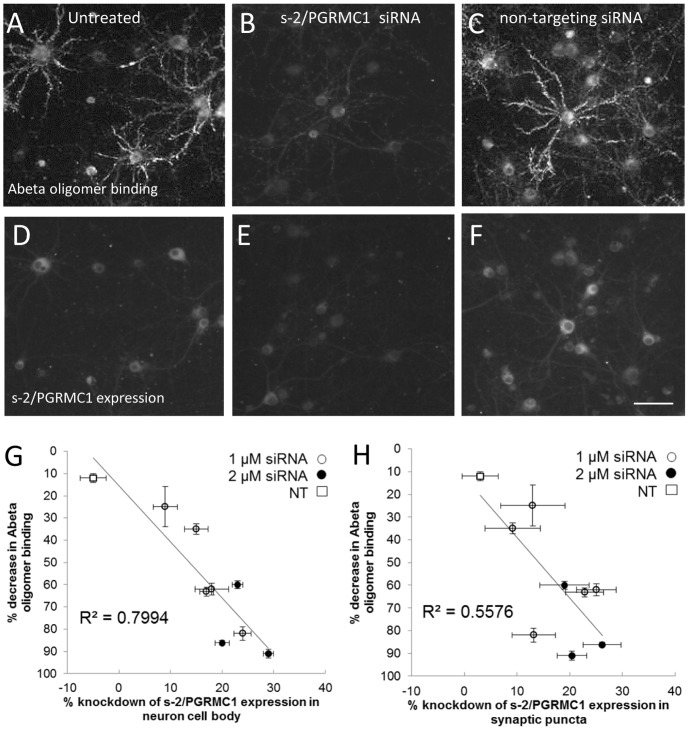
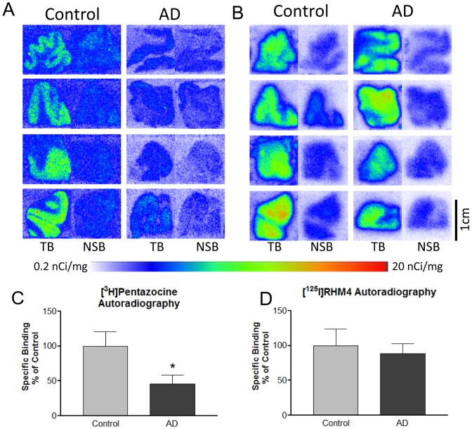
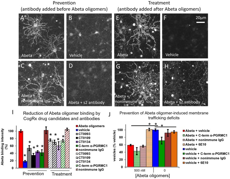
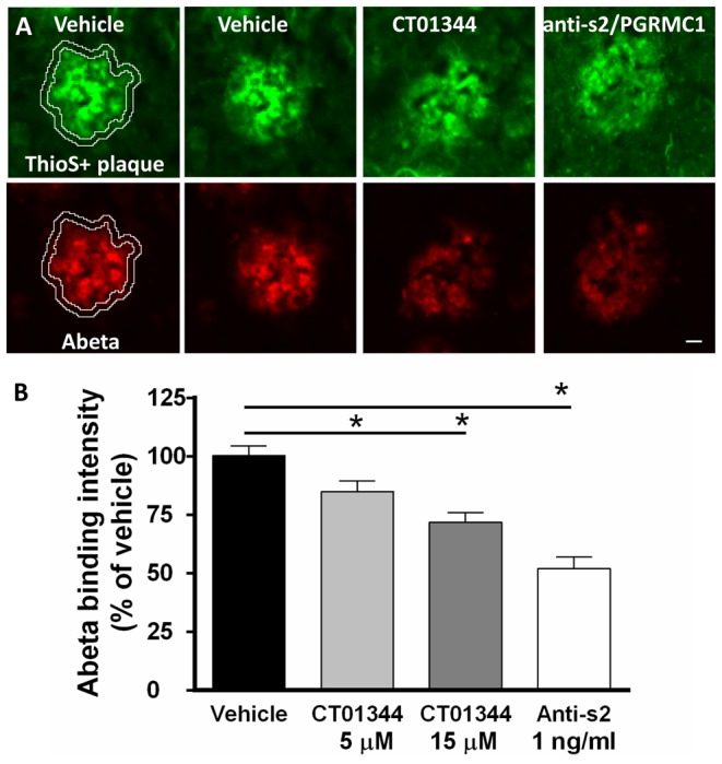
Amyloid beta (Abeta) 1-42 oligomers accumulate in brains of patients with Mild Cognitive Impairment (MCI) and disrupt synaptic plasticity processes that underlie memory formation. Synaptic binding of Abeta oligomers to several putative receptor proteins is reported to inhibit long-term potentiation, affect membrane trafficking and induce reversible spine loss in neurons, leading to impaired cognitive performance and ultimately to anterograde amnesia in the early stages of Alzheimer's disease (AD). We have identified a receptor not previously associated with AD that mediates the binding of Abeta oligomers to neurons, and describe novel therapeutic antagonists of this receptor capable of blocking Abeta toxic effects on synapses in vitro and cognitive deficits in vivo. Knockdown of sigma-2/PGRMC1 (progesterone receptor membrane component 1) protein expression in vitro using siRNA results in a highly correlated reduction in binding of exogenous Abeta oligomers to neurons of more than 90%. Expression of sigma-2/PGRMC1 is upregulated in vitro by treatment with Abeta oligomers, and is dysregulated in Alzheimer's disease patients' brain compared to age-matched, normal individuals. Specific, high affinity small molecule receptor antagonists and antibodies raised against specific regions on this receptor can displace synthetic Abeta oligomer binding to synaptic puncta in vitro and displace endogenous human AD patient oligomers from brain tissue sections in a dose-dependent manner. These receptor antagonists prevent and reverse the effects of Abeta oligomers on membrane trafficking and synapse loss in vitro and cognitive deficits in AD mouse models. These findings suggest sigma-2/PGRMC1 receptors mediate saturable oligomer binding to synaptic puncta on neurons and that brain penetrant, small molecules can displace endogenous and synthetic oligomers and improve cognitive deficits in AD models. We propose that sigma-2/PGRMC1 is a key mediator of the pathological effects of Abeta oligomers in AD and is a tractable target for small molecule disease-modifying therapeutics.


Conflict of interest statement

Competing Interests: NI, KM, CS, CR, RY, CL, GR, HS, and SC are employees or paid concultants of Cognition Therapeutics, Inc. HL and RC also own stock or stock options of Cognition Therapeutics.

Figures

Figure 1
Figure 1. Anti-Abeta compounds are ligands for sigma-2/PGRMC1 receptor.
A, CT0109, CT0093, CT01344 and CT01346 displace the fiduciary sigma-2 ligand [3H]-DTG from receptors on human B cell lines. B. Autoradiograms of 18.4 nM [125I]RHM-1 binding to human frontal cortex slices in the presence of 10, 100, 1000, 10,000 nM of CT0109 and CT0093, N = 4. Color bar under images show false coloring scale. [125I]RHM-1 displays specific saturable binding to human frontal cortex tissue as assessed by quantitative autoradiography in dose-response format (C) and as a Scatchard plot (D). E. Dose response curves for data obtained from autoradiograms in B. The Ki's for CT0109 and CT0093 at the [125I]RHM-1 binding site were 57±23 nM and 33±12 nM, respectively.
Figure 2
Figure 2. Sigma-2/PGRMC1 protein localizes to synaptic puncta on mature primary hippocampal cultures (21 days in vitro) and expression levels are positively correlated with Abeta oligomer binding.
sigma-2/PGRMC1 (A–D, red) is expressed at low levels in untreated cultures and is localized in cell bodies of neurons and glia, in neurite shafts, and adjacent to presynaptic puncta (A–D, synaptophysin  =  green) B. 66.7%±2.4 (average ± S.E.M., N = 110 neurons) of PGRMC1 positive puncta on neurons co-localize (yellow) with synaptophysin positive puncta. E–L. Positive correlation between sigma-2/PGRMC1 expression and Abeta oligomer binding in neurons (Abeta oligomers  = 400 nM, 1 hour treatment). E–H. Only one neuron (MAP2 positive arrow #1 in E–H) in this field is labeled with punctate Abeta oligomer binding (G), and exhibits elevated PGRMC1 expression (H, 3.3×10⁵ RFU) compared to surrounding neurons (#2 = 1.6×10⁵, #3 = 1.8×10⁵ RFU). I–L. Vehicle-treated cultures express a similar range of sigma-2/PGRMC1 expression in neurons (arrow #1 in I  = 2.62×10⁵, #2 = 1.21×10⁵ RFU). All scale bars  = 20 microns. M, Binning neurons according to their intensity of sigma-2/PGRMC1 immunofluorescence and graphing the average values for Abeta binding from each bin reveals a positive correlation between the intensity of Abeta oligomer binding to synaptic puncta and the expression of sigma-2/PGRMC1 in the cell body that is significant (Kruskal-Wallis, p<0.001). N. A similar analysis of sigma-2/PGRMC1 imunofluorescence in the synaptic puncta also shows a positive correlation with Abeta oligomer binding intensity to synaptic puncta (Kruskal-Wallis p<0.001).
Figure 3
Figure 3. Abeta oligomer treatment causes progressive upregulation of sigma-2/PGRMC1 expression.
Expression of sigma-2/PGRMC1 in the cell body of the entire culture cell population significantly increases with time of exposure to Abeta oligomers (A, C, E, 400 nM Abeta oligomers, *p = 0.05, ***p = 0.001, Student's t-test, N>1500 neurons and glia per condition) compared to vehicle-treated cultures (B, D, F). All scale bars  = 20 microns. G. Immunofluorescent intensity of sigma-2/PGRMC1 in Abeta oligomer-treated cells, relative to vehicle treatment, at 1 hr, 24 hr and 48 hr.
Figure 4
Figure 4. PGRMC1 mediates the binding of Abeta oligomers to neurons in vitro.
Co-immunolabeling for Abeta oligomer binding (A–C) and sigma-2/PGRMC1 expression (D–F) in the same field of view in hippocampal and cortical cultures (21DIV). Untreated neurons (A, D) exhibit Abeta oligomer binding to synaptic sites on neurites and low levels of sigma-2/PGRMC1 expression. In the presence of siRNA to sigma-2/PGRMC1, both Abeta oligomer binding and sigma-2/PGRMC1 expression are significantly reduced (B, E). Non-targeting siRNA (C, F) has no effect. G. H. Graphs of immunocytochemically detectable PGRMC1 protein expression associated with neuron cell bodies (G) and synaptic puncta (H), and Abeta oligomer binding to synapses for each of nine separate experiments (expressed as a percentage of untreated control culture values mean ± S.E.M.). siRNA-mediated reduction in PGRMC1 protein expression of up to 28% results in a corresponding decrease in Abeta oligomer binding by up to 91% (linear regression for PGRMC1 expression in neuronal cell bodies, r2 = 0.799, p = 0.0011; for PGRMC1 expression in synaptic puncta, r2 = 0.554, p = 0.02).
Figure 5
Figure 5. Sigma2/PGRMC1 receptor is present in human neocortex and is upregulated in AD.
A. Control receptor expression (unrelated receptor sigma-1 labeled with [3H]pentazocine 1 nM) declines in advanced AD patient frontal cortex sections vs. age-matched cognitively normal individuals (N = 4, *p = 0.0375, Student's t-test). B, adjacent sections from the same individuals labeled with [125I] sigma-2 antagonist RHM-4 (0.2 nM) reveal sigma-2 receptor expression is not significantly changed in the disease state vs. normal individuals (N = 4). TB  =  Total binding. NSB  =  non-specific binding in presence of 10 fold excess cold ligand. C, D, Quantified values for binding in images shown in A and B expressed as the percent of specific binding in controls.
Figure 6
Figure 6. C-terminal antibodies directed against the C-terminus of PGRMC1 prevent (A–D) and displace (E–H) Abeta oligomer binding to neurons and glia.
Abeta oligomers bind to a subset of neurons and glia in mature hippocampal primary neurons 21DIV (A, E, red bar in I) compared to vehicle-treated (no Abeta) cultures (B, F, blue bar in I). Graphs in I are average of 3 experiments (avg. intensity of Abeta oligomer puncta + S.E.M., expressed as a percentage of Abeta oligomer-treated condition, difference in binding intensity vs. Abeta oligomer condition *p<0.05, Student's t-test). Abeta oligomer binding to cultured neurons is significantly reduced in the presence of C-terminal antibody to sigma-2/PGRMC1 regardless of whether it is added before (D, green bar in I [prevention], 58% reduction) or after (H, green hatched bar in I [treatment], 26% reduction) oligomers. This suggests that oligomers are competitively displaced from receptors at synaptic sites. Non-immune IgG (C, G and maroon bars in I) and an N-terminal antibody to sigma-2/PGRMC1 (data not shown) cannot reduce oligomer binding under either condition. J Effects of antibodies on membrane trafficking rate in the presence or absence of Abeta oligomers (expressed as a percentage of vehicle-treated in the absence of Abeta, difference in trafficking rate vs. Abeta oligomer- or vehicle-treated condition *p<0.05, Student's t-test). The C-terminal antibody directed against amino acids 185–195 in sigma-2/PGRMC1 does not rescue oligomer-induced deficits, but induces trafficking deficits on its own in the absence of Abeta oligomers, pointing to a critical role of this protein in normal membrane trafficking.
Figure 7
Figure 7. Antibodies and compounds directed against sigma-2/PGRMC1 dose-dependently displace endogenous human Abeta oligomers from specific tissue locations in Alzheimer's brain neocortical tissue sections.
A, Abeta oligomers located in a 2 micron halo surrounding compact thioflavin-S positive plaques is displaced from frozen postmortem human AD brain tissue sections (N = 7 patients) by C-terminal sigma-2/PGRMC1 antibody and receptor antagonist CT01344 (B) in a dose-dependent manner compared to vehicle-treated brain sections from the same individual (Spearman's rank order, rho  = −0.492, p = 0.038). All scale bars  = 20 microns. See Fig. S4 for details on quantification method.
Figure 8
Figure 8. CogRx sigma-2/PGRMC1-selective small molecules are functional antagonists.
A, B Sigma-2/PGRMC1 agonist siramesine causes dose-dependent activation of caspase 3 in primary neuronal cultures (A) and in SKOV-3 human ovarian cancer cells (B) but sigma-2/PGRMC1 antagonists RHM-1, CT0109 and CT0093 do not. C, D Sigma-2/PGRMC1 agonists siramesine, WC-26 and SV-119 cause dose-dependent cell death in primary hippocampal/cortical cultures (C) and in SKOV-3 human ovarian cancer cells (D) but sigma-2/PGRMC1 antagonists RHM-1, CT0109 and CT0093 do not, except at very high concentrations (>100 µM). (E) Treatment of cultures of hippocampal and cortical cells with 20 to 80 µM SV-119 for 24 hours induced the activation of caspase 3/7 (*p<0.05 by 2-tailed Student's t-test compared to control). Co-treatment of cultures with 40 µM CT0109 or CT0093 did not increase caspase activity and blocked the activation by the agonist SV-119.

References

    1. Klein WL, Krafft G, Finch CE (2001) Targeting small Abeta oligomers: the solution to an Alzheimer's disease conundrum. Trends Neurosci 24: 219–224. - PubMed
    1. Mucke L, Selkoe DJ (2012) Neurotoxicity of Amyloid beta-Protein: Synaptic and Network Dysfunction. Cold Spring Harb Perspect Med 2: a006338. - PMC - PubMed
    1. Bao F, Wicklund L, Lacor PN, Klein WL, Nordberg A, et al. (2012) Different beta-amyloid oligomer assemblies in Alzheimer brains correlate with age of disease onset and impaired cholinergic activity. Neurobiol Aging 33: 813 –813. - PubMed
    1. Haes AJ, Chang L, Klein WL, Van Duyne RP (2005) Detection of a biomarker for Alzheimer's disease from synthetic and clinical samples using a nanoscale optical biosensor. J Am Chem Soc 127: 2264–2271. - PubMed
    1. Tomic JL, Pensalfini A, Head E, Glabe CG (2009) Soluble fibrillar oligomer levels are elevated in Alzheimer's disease brain and correlate with cognitive dysfunction. Neurobiol Dis 35: 352–358. - PMC - PubMed

Publication types

MeSH terms