. 2014 Nov 10;15(11):20555-71.
doi: 10.3390/ijms151120555.

Depletion of C3orf1/TIMMDC1 inhibits migration and proliferation in 95D lung carcinoma cells

Affiliations

Depletion of C3orf1/TIMMDC1 inhibits migration and proliferation in 95D lung carcinoma cells

Huiling Wu et al. Int J Mol Sci. .

Abstract

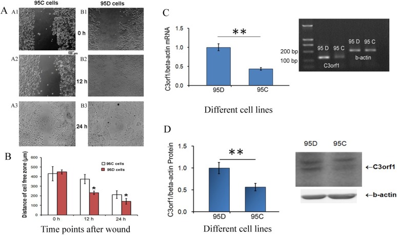
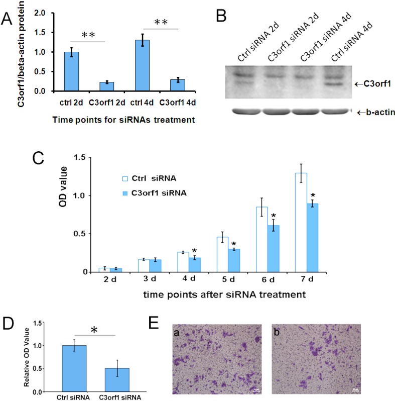
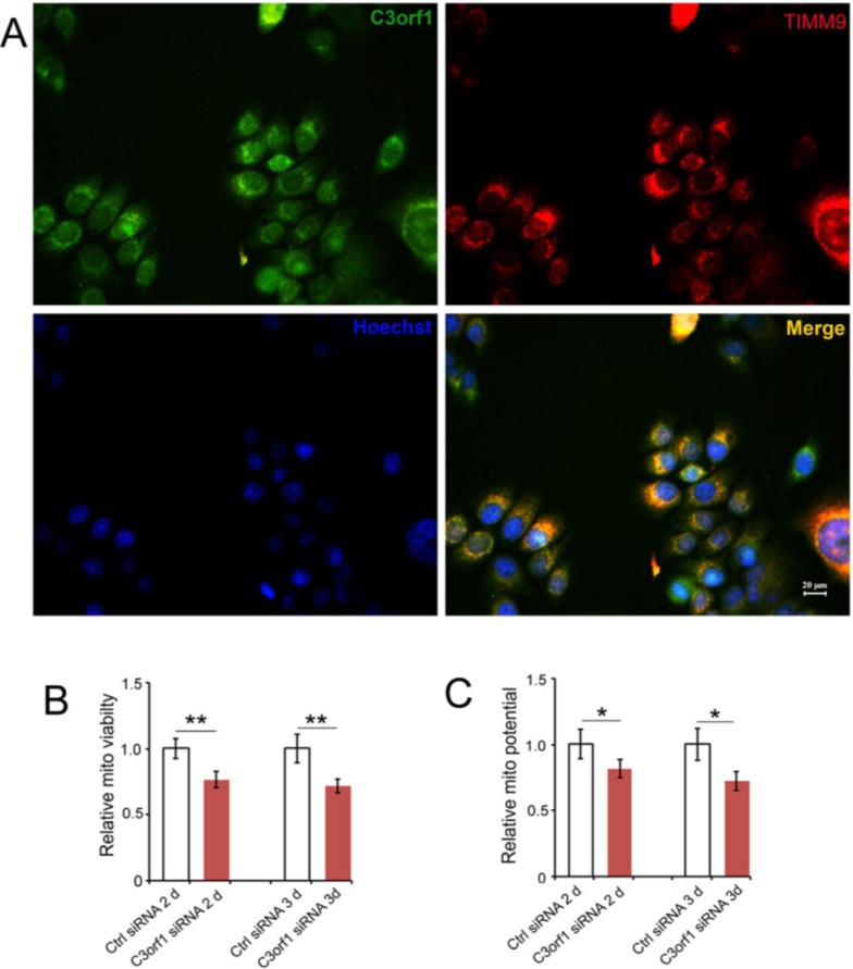
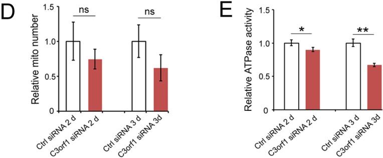
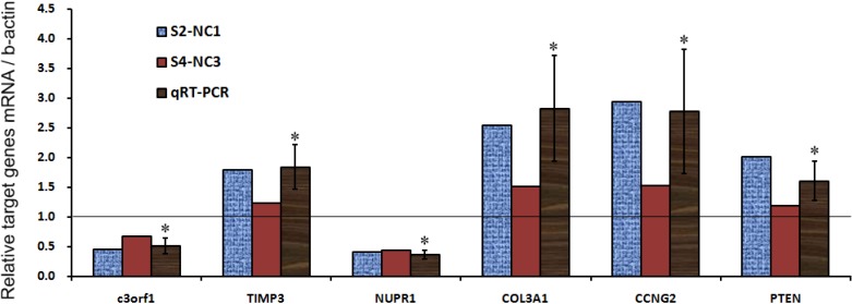
In our previous study, we identified an association of high expression of c3orf1, also known as TIMMDC1 (translocase of inner mitochondrial membrane domain-containing protein 1), with metastatic characteristics in lung carcinoma cells. To investigate the preliminary function and mechanism of this mitochondrial protein, we depleted C3orf1 expression by introducing siRNA into 95D lung carcinoma cells. We demonstrated that C3orf1 depletion significantly suppressed 95D cell growth and migration. We confirmed C3orf1 localization in the inner mitochondrial membrane and showed that mitochondrial viability, membrane potential, and ATPase activity were remarkably reduced upon depletion of C3orf1. Microarray data indicated that genes involved in regulation of cell death, migration, and cell-cycle arrest were significantly altered after C3orf1 depletion for 48 h. The expression of genes involved in focal adhesion, ECM-receptor interaction, and p53-signaling pathways were notably altered. Furthermore, cell-cycle arrest genes such as CCNG2 and PTEN as well as genes involved in cell migration inhibition, such as TIMP3 and COL3A1, were upregulated after C3orf1 depletion in 95D cells. Concurrently, expression of the migration-promoting gene NUPR1 was markedly reduced, as confirmed by real-time PCR. We conclude that C3orf1 is critical for mitochondrial function, migration, and proliferation in 95D lung carcinoma cells. Depletion of C3orf1 inhibited cell migration and cell proliferation in association with upregulation of genes involved in cell-cycle arrest and cell migration inhibition. These results suggest that C3orf1 (TIMMDC1) may be a viable treatment target for lung carcinoma, and that further study of the role of this protein in lung carcinoma pathogenesis is justified.


Figures

Figure 1
Figure 1
Expression of the C3orf1 gene is high in migratory 95D lung carcinoma cells. (A) Results of wound-healing assays in 95C and 95D cells. Panel A1–A3, representative images of scratch-wounded 95C cells at 0, 12, and 24 h. Panel B1–B3, representative images of scratch-wounded 95D cells at 0, 12, and 24 h. Scale bar = 100 µm; (B) Statistical results of wound healing assays in 95C and 95D cells. Measurements of the distance of wounded cell-free space at the indicated time points. Data are represented as mean ± SD; n = 3; * p < 0.05; (C) Results of real-time quantitative PCR measuring the relative c3orf1 mRNA level in lung carcinoma 95D and 95C cells. β-actin was used as an internal control. Data are represented as mean ± SD. The data were normalized (i.e., the average expression level of c3orf1 in 95D cells was set to 1, and the average expression level of c3orf1 in 95C cells was calculated in relation to this averaged value); n = 4, ** p < 0.01. The insert panel is a representative image of the semi-quantitative RT-PCR gel; and (D) Results of Western blotting analysis used to detect the relative C3orf1 protein level in lung carcinoma 95D and 95C cells. β-actin was used as the internal control. Data are represented as mean ± SD. The data were normalized as described in Figure 1C; n = 3, ** p < 0.01. The insert panel shows representative images of Western blotting results.
Figure 2
Figure 2
Depletion of C3orf1 in 95D cells inhibits cell proliferation and migration. (A) The efficiency of different siRNAs was evaluated by Western blotting after siRNA transfection in 95D cells for 2 days (2d) and 4 days (4d). β-actin was used as an internal control. Data are represented as mean ± SD. The data were normalized as described above, and the average expression level of C3orf1 protein in 95D cells treated with ctrl siRNA for 2 days (2d) was set to 1; ** p < 0.01, n = 3; (B) Representative images of Western blotting results; (C) The cell proliferation curve indicating that depleting C3orf1 expression suppressed the proliferation of 95D cells significantly between day 4 (4d) and day 7 (7d). The data are represented as mean ± SD; * p < 0.05, n = 3; (D) Trans-well assays indicating that depleting C3orf1 expression decreased cell migration. Data are represented as mean ± SD. The data were normalized, and the average number of migrated 95D cells treated with ctrl siRNA was set to 1; * p < 0.05, n = 4; and (E) Representative images of stained trans-well assay membranes. Scale bar = 50 µm.
Figure 3
Figure 3
C3orf1 localizes to the inner mitochondrial membrane of 95D cells and C3orf1 knockdown affects mitochondria-related functions; (A) Representative images of immunostaining with anti-C3orf1 antibody (in green) and anti-Timm9 antibody (in red). The nucleus was labeled with Hoechst33342 dye (in blue). Scales bar = 20 µm; (B) Results of mitochondrial viability assays on 95D cells subjected to control or c3orf1 siRNA treatment for 2 days (2d) and 3 days (3d). Data are represented as mean ± SD. The data were normalized as described above, and the average mitochondrial viability of 95D cells treated with ctrl siRNA was set to 1; ** p < 0.01, n = 3; (C) Results of mitochondrial potential assays on 95D cells subjected to control or c3orf1 siRNA treatment for 2 days (2d) and 3 days (3d). Data are represented as mean ± SD. The data were normalized, and the average mitochondrial potential of 95D cells treated with ctrl siRNA was set to 1; * p < 0.05, n = 3; (D) Results of mitochondrial number assays in 95D cells subjected to control or c3orf1 siRNA treatment for 2 days (2d) and 3 days (3d). Data are represented as mean ± SD. The data were normalized, and the average mitochondrial number of 95D cells treated with ctrl siRNA was set to 1; n = 3; ns = no significant change in mitochondrial numbers between cells treated with control siRNA or c3orf1 siRNA; and (E) Results of ATPase activity assays on 95D cells subjected to control or c3orf1 siRNA treatment for 2 days (2d) and 3 days (3d). Data are represented as mean ± SD. The data were normalized, and the average ATPase activity of 95D cells treated with ctrl siRNA was set to 1; * p < 0.05, ** p < 0.01, n = 3.
Figure 3
Figure 3
C3orf1 localizes to the inner mitochondrial membrane of 95D cells and C3orf1 knockdown affects mitochondria-related functions; (A) Representative images of immunostaining with anti-C3orf1 antibody (in green) and anti-Timm9 antibody (in red). The nucleus was labeled with Hoechst33342 dye (in blue). Scales bar = 20 µm; (B) Results of mitochondrial viability assays on 95D cells subjected to control or c3orf1 siRNA treatment for 2 days (2d) and 3 days (3d). Data are represented as mean ± SD. The data were normalized as described above, and the average mitochondrial viability of 95D cells treated with ctrl siRNA was set to 1; ** p < 0.01, n = 3; (C) Results of mitochondrial potential assays on 95D cells subjected to control or c3orf1 siRNA treatment for 2 days (2d) and 3 days (3d). Data are represented as mean ± SD. The data were normalized, and the average mitochondrial potential of 95D cells treated with ctrl siRNA was set to 1; * p < 0.05, n = 3; (D) Results of mitochondrial number assays in 95D cells subjected to control or c3orf1 siRNA treatment for 2 days (2d) and 3 days (3d). Data are represented as mean ± SD. The data were normalized, and the average mitochondrial number of 95D cells treated with ctrl siRNA was set to 1; n = 3; ns = no significant change in mitochondrial numbers between cells treated with control siRNA or c3orf1 siRNA; and (E) Results of ATPase activity assays on 95D cells subjected to control or c3orf1 siRNA treatment for 2 days (2d) and 3 days (3d). Data are represented as mean ± SD. The data were normalized, and the average ATPase activity of 95D cells treated with ctrl siRNA was set to 1; * p < 0.05, ** p < 0.01, n = 3.
Figure 4
Figure 4
Alteration in the gene expression profile of C3orf1-depleted cells. Graphic representation of real-time PCR results depicting the expression of selected genes; * p < 0.05.

References

    1. Siegel R., Ma J.M., Zou Z.H., Jemal A. Cancer Statistics, 2014. CA Cancer J. Clin. 2014;64:9–29. - PubMed
    1. Chen W.Q., Zheng R.S., Zhang S.W., Zhao P., Zeng H.M., Zou X.N., He J. Annual report on status of cancer in China, 2010. Chin. J. Cancer Res. 2014;26:48–58. - PMC - PubMed
    1. Gong H.C., Wang S., Mayer G., Chen G., Leesman G., Singh S., Beer D.G. Signatures of drug sensitivity in nonsmall cell lung cancer. Int. J. Proteomics. 2011;2011:215496. doi: 10.1155/2011/215496. - DOI - PMC - PubMed
    1. Ellis P.M., Al-Saleh K. Multitargeted anti-angiogenic agents and NSCLC: Clinical update and future directions. Crit. Rev. Oncol. Hematol. 2012;84:47–58. - PubMed
    1. Shepherd F.A., Douillard J.Y., Blumenschein G.R., Jr. Immunotherapy for non-small cell lung cancer: Novel approaches to improve patient outcome. J. Thorac. Oncol. 2011;6:1763–1773. - PubMed

Publication types

MeSH terms

Substances