. 2015 Jan 9;290(2):762-74.
doi: 10.1074/jbc.M114.608497. Epub 2014 Nov 12.

Selective inhibition of mutant isocitrate dehydrogenase 1 (IDH1) via disruption of a metal binding network by an allosteric small molecule

Affiliations

Selective inhibition of mutant isocitrate dehydrogenase 1 (IDH1) via disruption of a metal binding network by an allosteric small molecule

Gejing Deng et al. J Biol Chem. .

Abstract

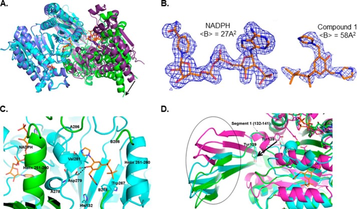
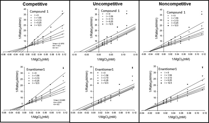
Cancer-associated point mutations in isocitrate dehydrogenase 1 and 2 (IDH1 and IDH2) confer a neomorphic enzymatic activity: the reduction of α-ketoglutarate to d-2-hydroxyglutaric acid, which is proposed to act as an oncogenic metabolite by inducing hypermethylation of histones and DNA. Although selective inhibitors of mutant IDH1 and IDH2 have been identified and are currently under investigation as potential cancer therapeutics, the mechanistic basis for their selectivity is not yet well understood. A high throughput screen for selective inhibitors of IDH1 bearing the oncogenic mutation R132H identified compound 1, a bis-imidazole phenol that inhibits d-2-hydroxyglutaric acid production in cells. We investigated the mode of inhibition of compound 1 and a previously published IDH1 mutant inhibitor with a different chemical scaffold. Steady-state kinetics and biophysical studies show that both of these compounds selectively inhibit mutant IDH1 by binding to an allosteric site and that inhibition is competitive with respect to Mg(2+). A crystal structure of compound 1 complexed with R132H IDH1 indicates that the inhibitor binds at the dimer interface and makes direct contact with a residue involved in binding of the catalytically essential divalent cation. These results show that targeting a divalent cation binding residue can enable selective inhibition of mutant IDH1 and suggest that differences in magnesium binding between wild-type and mutant enzymes may contribute to the inhibitors' selectivity for the mutant enzyme.

Keywords: Anticancer Drug; Cancer Metabolism; Drug Action; Drug Discovery; Enzyme Inhibitor; IDH1 Inhibitor; Mechanism of Inhibition; Metalloenzyme.


Figures

FIGURE 1.
FIGURE 1.
Time-dependent inhibition of R132H IDH1 by compounds 1 and 2. Compounds were preincubated with the enzyme for the time indicated, and IC50 values were determined as described under “Experimental Procedures.”
FIGURE 2.
FIGURE 2.
Compounds 1 and 2 inhibit 2HG production in R132H IDH1-expressing HEK-293 cells. Cells were treated with compound or DMSO (final DMSO concentration, 0.5%) for 24 h before 20 μl of cell culture supernatant was harvested for 2HG quantitation by LC-MS/MS. Percentage of 2HG inhibition: %I = 100 × (1 − (amount of 2HG in compound treated sample/amount of 2HG in DMSO-treated sample)).
FIGURE 3.
FIGURE 3.
Compound 1 inhibits R132H IDH1 noncompetitively with respect to NADPH and αKG, respectively. Alternative inhibition models were fitted to steady-state inhibition data for compound 1. Inhibition data with respect to NADPH are illustrated with Michaelis-Menten plots (top row), whereas data with respect to αKG are illustrated with Lineweaver-Burk plots (bottom row). The fits to the noncompetitive model (far-right panel) are the best, as judged by comparison of AICc. The unit of inhibitor concentrations shown in the insets is micromolar.
FIGURE 4.
FIGURE 4.
A, compounds 1 and 2 bind to both free and NADPH-bound R132H IDH1 enzyme forms and bind selectively to one enantiomer of compound 2. SPR binding isotherms were measured for compound 1 (top panel), compound 2 (middle panel), and the two enantiomers of compound 2 (bottom panel). Saturable binding of only the active enantiomer (enantiomer 1) but not the inactive enantiomer (enantiomer 2) indicates that inhibitor binding to R132H IDH1 immobilized on SPR sensor chip is due to a specific interaction. The data were fitted to a steady-state affinity model. For the purpose of comparison, biochemical IC50 and cellular IC50 (2HG inhibition) of each compound are shown on the top of each SPR binding isotherm. B, immobilization of R132H IDH1 onto the SPR chip surface does not compromise binding of its native ligands NADPH and NADP+. Binding and dissociation time traces (left panels) are fitted to a 1:1 kinetic binding model (original data and fits are shown in colors and in black, respectively), and binding isotherms obtained from steady-state response levels (right panels); response units were determined at 4 s before the end of each compound injection) are independently fitted to the Langmuir equation. The KD values calculated based on kon and koff from the fits of the binding kinetics are consistent with the values obtained by fitting the steady-state binding data.
FIGURE 5.
FIGURE 5.
X-ray structure of compound 1 with homodimeric R132H IDH1. A, superimposition of structures of R132H IDH1/NADP+ dimer (blue/violet) and R132H IDH1/NADP+/compound 1 dimer (cyan/green), with the A protomers aligned. A large movement of the B protomer can be seen upon binding of compound 1 (black arrow). B, electron density maps of NADPH and compound 1. C, close-up of the dimer interface in the R132H IDH1/NADPH/compound 1 structure (A protomer in cyan and B protomer in green). D, the conformation of the clasp domain (circled) and segment 1 in the compound 1-bound structure and 3INM. Segment 1 (residue 132–141) in the compound 1-bound structure is disordered (green and cyan for protomer A and protomer B, respectively). Tyr139, a residue believed to play a vital role in catalysis, is no longer oriented toward the active site as it is in the substrate-bound structure (magenta).
FIGURE 6.
FIGURE 6.
Both compounds 1 and 2 inhibit R132H IDH1 competitively with respect to Mg2+. Alternative inhibition models were fitted to steady-state inhibition data and are illustrated with Lineweaver-Burk plots. The fits to the competitive model (left panel) are the best, as judged by comparison of AICc. The unit of inhibitor concentrations shown in the insets is nanomolar.
FIGURE 7.
FIGURE 7.
Proposed kinetic mechanism for inhibition of the reductive reaction of R132H IDH1. A, NADPH; B, αKG; M, Mg2+; I, compound 1 or compound 2; P, 2HG; Q, NADP+. Species in black have either been detected directly via SPR or are required to accommodate steady-state kinetics results; the involvement of species in gray is neither required nor ruled out by experimental results to date.

References

    1. Tennant D. A., Durán R. V., Gottlieb E. (2010) Targeting metabolic transformation for cancer therapy. Nat. Rev. Cancer 10, 267–277 - PubMed
    1. Levine A. J., Puzio-Kuter A. M. (2010) The control of the metabolic switch in cancers by oncogenes and tumor suppressor genes. Science 330, 1340–1344 - PubMed
    1. Reitman Z. J., Yan H. (2010) Isocitrate dehydrogenase 1 and 2 mutations in cancer: alterations at a crossroads of cellular metabolism. J. Natl. Cancer Inst. 102, 932–941 - PMC - PubMed
    1. Dang L., Jin S., Su S. M. (2010) IDH mutations in glioma and acute myeloid leukemia. Trends Mol. Med. 16, 387–397 - PubMed
    1. Geisbrecht B. V., Gould S. J. (1999) The human PICD gene encodes a cytoplasmic and peroxisomal NADP+-dependent isocitrate dehydrogenase. J. Biol. Chem. 274, 30527–30533 - PubMed

MeSH terms

LinkOut - more resources