. 2015 Jan 2;290(1):478-91.
doi: 10.1074/jbc.M114.593632. Epub 2014 Nov 12.

Conformational dynamics of the focal adhesion targeting domain control specific functions of focal adhesion kinase in cells

Affiliations

Conformational dynamics of the focal adhesion targeting domain control specific functions of focal adhesion kinase in cells

Gress Kadaré et al. J Biol Chem. .

Abstract

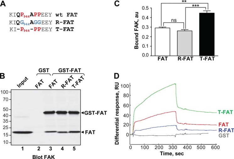
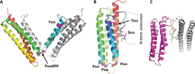
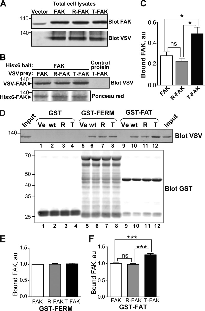
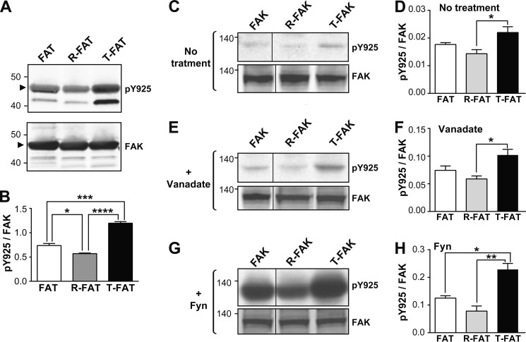
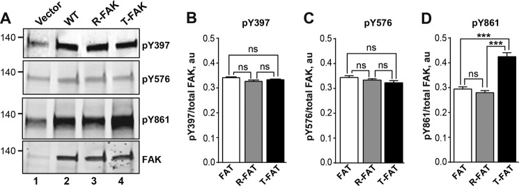
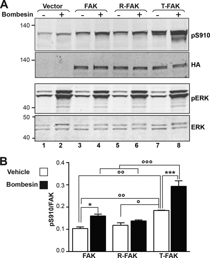
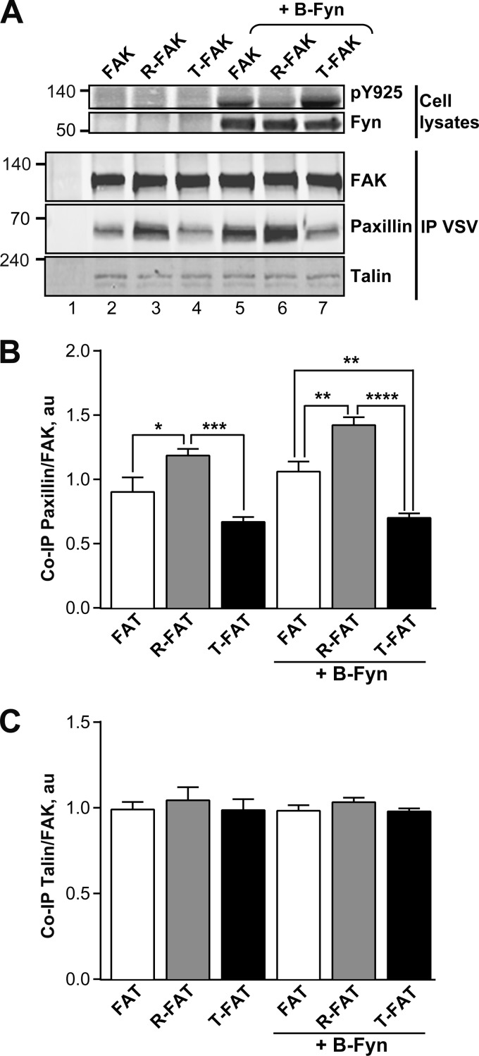
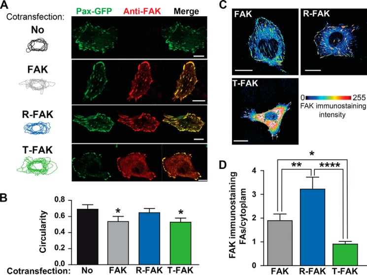
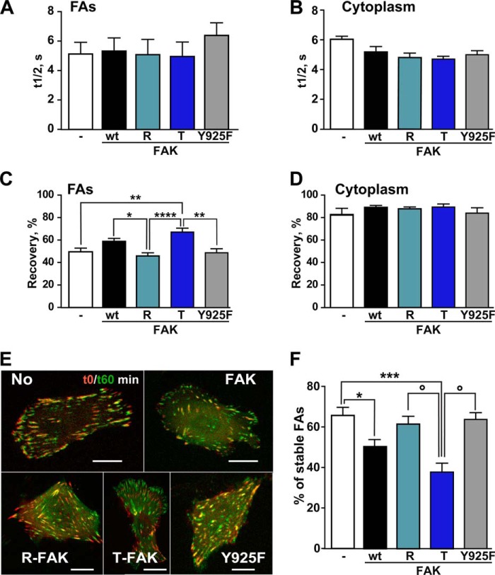
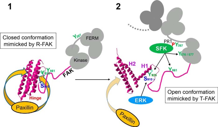
Focal adhesion (FA) kinase (FAK) regulates cell survival and motility by transducing signals from membrane receptors. The C-terminal FA targeting (FAT) domain of FAK fulfils multiple functions, including recruitment to FAs through paxillin binding. Phosphorylation of FAT on Tyr(925) facilitates FA disassembly and connects to the MAPK pathway through Grb2 association, but requires dissociation of the first helix (H1) of the four-helix bundle of FAT. We investigated the importance of H1 opening in cells by comparing the properties of FAK molecules containing wild-type or mutated FAT with impaired or facilitated H1 openings. These mutations did not alter the activation of FAK, but selectively affected its cellular functions, including self-association, Tyr(925) phosphorylation, paxillin binding, and FA targeting and turnover. Phosphorylation of Tyr(861), located between the kinase and FAT domains, was also enhanced by the mutation that opened the FAT bundle. Similarly phosphorylation of Ser(910) by ERK in response to bombesin was increased by FAT opening. Although FAK molecules with the mutation favoring FAT opening were poorly recruited at FAs, they efficiently restored FA turnover and cell shape in FAK-deficient cells. In contrast, the mutation preventing H1 opening markedly impaired FAK function. Our data support the biological importance of conformational dynamics of the FAT domain and its functional interactions with other parts of the molecule.

Keywords: Conformational Change; Focal Adhesions; Protein Kinase; Protein Structure; Tyrosine-Protein Kinase (Tyrosine Kinase).


Figures

FIGURE 1.
FIGURE 1.
Mutations in the H1-H2 hinge of FAT alter its self-association. A, mutations designed to relax (R) or increase (T) the tension in the HI-H2 FAT hinge region. B, pull-down assays with immobilized GST-FAT or GST and purified FAT domain: WT-FAT (lanes 2 and 3), R-FAT (lane 4), and T-FAT (lane 5). Input (lane 1): 1 μg of soluble FAT equivalent to the total amount used in the pull-down assays. Molecular mass markers positions are indicated in kDa. C, quantification of bound FAT in three independent pull-down experiments (mean ± S.E., a.u., arbitrary units). One-way ANOVA, F2,6 = 37.7, p = 0.0004, Tukey's test **, p < 0.01; ***, p < 0.001; ns, not significant. D, representative surface plasmon resonance sensorgrams showing the binding curves of purified FAT domains to a sensor chip covalently coated with purified GST-FAT. RU, resonance units. Differential response was obtained by subtracting the signal in the blank channel from that in the experimental channel. Estimated Kd: FAT, 23 ± 8.6 μm; T-FAT, 1.3 ± 0.8 μm; R-FAT, 24 ± 5.5 μm (mean ± S.E., n = 3).
FIGURE 2.
FIGURE 2.
Structural analysis of FAT predicts several possible modes of dimerization. A, crystallographic model for the H1-swapped FAT dimer (PDB accession 1K04). One protomer is shown in gray; the other one is color-ramped from the N terminus (blue) to the C terminus (red). B, interaction in cis between the N-terminal extension (residues 908–915) and FAT H1/H4 observed in the FAT(892–1052) crystal structure determined to a resolution of 2.6 Å for this study (Table 1, PDB accession 3S9O, molecule A is shown). The secondary structure representation is color-ramped as in A. Side chains of relevant residues are shown. C, crystallographic model for a potential FAT-FAT interaction via swapping of the N-terminal extensions (residues 908–915). This arrangement is also taken from PDB accession 3S9O, molecule C, and symmetry-related molecule C.
FIGURE 3.
FIGURE 3.
T-FAT mutation increases full-length FAK dimerization and interaction with FAT. A–C, mutant and WT FAK proteins with N-terminal VSV tags were expressed by transfection in COS7 cells. A, immunoblotting of FAK in 20% of the amount of cell lysates used for each pull-down. Molecular mass marker positions are in kDa. B, cell lysates were loaded onto Ni2+ beads coated with recombinant His6-FAK (left lanes) or a His6-tagged 65-kDa unrelated protein (right lane). After extensive washing, bound proteins were eluted and the presence of VSV-tagged protein was assessed by immunoblotting. Ponceau red staining of the membrane shows equivalent amounts of His6-FAK bait. C, quantification of the pull-down experiment performed in triplicate (mean ± S.E., au, arbitrary units). One-way ANOVA, F(2,6) = 9.8, p = 0.013, Tukey's test; *, p < 0.05; ns, not significant. In these experiments, full-length FAK interactions are likely to involve FERM-FERM or FAT-FERM interactions, in addition to the FAT-FAT interactions. D, COS7 cell lysates containing identical amounts of VSV-tagged mutant or WT FAK proteins were added to beads coated with GST (lanes 1–4), GST-FERM (lanes 5–8), and GST-FAT (lanes 9–12). After washing and elution, bound FAK was visualized by immunoblotting with VSV antibodies. Equivalent quantities of the three different baits used (GST, GST-FERM, and GST-FAT) were revealed by immunoblotting with a GST antibody. Input (20% of the lysate amount used in the pull-down) is shown on either side of the VSV blot. E and F, quantification of the amount of VSV-FAK retained by GST-FERM (B) and GST-FAT (C) in three independent experiments (mean ± S.E., au, arbitrary units). One-way ANOVA GST-FERM, no significant difference, GST-FAT, F(2,6) = 43, p < 0.001, Tukey's test; ***, p < 0.001; ns, not significant. The T mutation in full-length FAK did not modify its binding to GST-FERM but increased its interaction with GST-FAT. A, B, and D, molecular mass marker positions are indicated in kDa.
FIGURE 4.
FIGURE 4.
Mutations of the FAT H1-H2 hinge alter phosphorylation of Tyr925. A, purified GST-tagged WT FAT domain (lane 1), R-FAT (lane 2, R-FAT), and T-FAT (lane 3) were incubated with Src (0.1 μg/ml) in the presence of ATP for 15 min at 30 °C. Immunoblotting was carried out with Tyr925 phospho-specific antibody (pY925, upper panel) or a FAK C-terminal antibody (FAK, lower panel). The GST-FAT position is indicated by an arrowhead. Note that phosphorylation of a breakdown product with a lower molecular weight was also affected in the same direction as full-length FAT-GST by the hinge mutations. B, quantification of three experiments for Tyr(P)925 as in A, corrected for the amount of FAK (mean ± S.E.). One-way ANOVA, F(2,6) = 105, p < 0.0001, Tukey's test; *, p < 0.05; ***, p < 0.001; ****, p < 0.0001. C, COS7 cells were transfected with WT or mutated VSV-tagged FAK and pY925 and FAK were analyzed by immunoblotting. D, quantification of results in C. One-way ANOVA, F(2,6) = 6.43, p < 0.05. E and F, same as in C and D, except that cells were treated for 16 h with 50 μm orthovanadate before lysis. One-way ANOVA, F(2,9) = 7.28, p < 0.05. G and H, same as in C and D, except that FAK was cotransfected with B-Fyn. One-way ANOVA, F(2,6) = 19.9, p = 0.002. D, F, and H, Tukey's test, *, p < 0.05; **, p < 0.01. A, C, E, and G, molecular mass markers positions are indicated in kDa. C, E, and G) the samples were run on the same gel and blot but intervening lanes were deleted as indicated by a vertical line.
FIGURE 5.
FIGURE 5.
Mutation of the FAT H1-H2 hinge alters phosphorylation of Tyr861 but not Tyr397 or Tyr576. A, COS7 cells were transfected with WT FAK, R-FAK, or T-FAK. Representative blots show phosphorylation of Tyr397, Tyr576, and Tyr861 using phospho-specific antibodies (pY397, pY576, and pY861, respectively). Levels of FAK protein were assessed with 4.47 monoclonal antibody (FAK). Molecular mass markers positions are indicated in kDa. B–D, quantification of three experiments as in A, for Tyr(P)397 (B), Tyr(P)576 (C), and Tyr(P)861 (D), corrected for the amount of FAK (mean ± S.E.). Statistical analysis, one-way ANOVA: Tyr(P)397, F(2,6) = 3.65, p = 0.09; Tyr(P)576, F(2,6) = 2.82, p = 0.14; Tyr(P)861, F(6,2) = 48.1, p = 0.0002, Tukey's post hoc test; ***, p < 0.001; ns, not significant.
FIGURE 6.
FIGURE 6.
Effects of the FAT hinge mutation on Ser910 phosphorylation. A, COS7 cells transfected with HA-tagged WT FAK, R-FAK, or T-FAK were serum deprived for 30 min and then treated for another 30 min with 10 nm bombesin before cell lysis. Phosphorylation of Ser910 and levels of transfected FAK expression were analyzed by immunoblotting with phospho-specific Ser(P)910 (pS910) and HA antibodies (upper panels). In parallel, diphospho-44/42 ERK (pERK) and total ERK were monitored with the corresponding antibodies (lower panels). Molecular mass marker positions are indicated in kDa. B, quantification of three experiments for Ser(P)910 as in A, corrected for the amount of FAK (mean ± S.E.). Two-way ANOVA, interaction; F(2,8) = 5.96, p = 0.026, treatment; F(1,4) = 80.9, p = 0.0008, mutation; F(2,8) = 44.7, p < 0.0001. Sidak's multiple comparisons test, asterisks; ± bombesin, ○; comparisons between FAK constructs, 1 symbol, p < 0.05; 2 symbols, p < 0.01; 3 symbols p < 0.001.
FIGURE 7.
FIGURE 7.
Mutations of the FAT H1-H2 hinge alter interaction of FAK with paxillin but not talin. A, COS7 cells were transfected with VSV-tagged WT FAK, R-FAK, or T-FAK, without or with B-Fyn cotransfection, as indicated. Cells were lysed 24 h later and FAK immunoprecipitated with anti-VSV antibodies (IP VSV). Phosphorylated Tyr925 was determined in total cell lysates (pY925, upper panel). Expression of B-Fyn was detected by immunoblotting. Immune precipitates were probed with FAK, paxillin, and talin antibodies as indicated (lower panels). Mass markers positions are indicated in kDa. B, quantification of the co-immunoprecipitation of paxillin with FAK as in A expressed as a ratio of paxillin/FAK, a.u., arbitrary units. Statistical analysis, one-way ANOVA: F(5,18) = 18.09, p < 0.0001, Tukey's post hoc test; *, p < 0.05; **, p < 0.01; ***, p < 0.001; ****, p < 0.0001. C, quantification of the co-immunoprecipitation of talin with FAK as in A expressed as a ratio of talin/FAK, a.u., arbitrary units. One-way ANOVA: F(5,18) = 0.35, p = 0.87. B and C, data are mean ± S.E.
FIGURE 8.
FIGURE 8.
Intracellular localization of FAT H1-H2 hinge mutants. A, FAK−/− fibroblasts were co-transfected with paxillin-GFP without (No) or with various VSV-tagged FAK constructs (WT FAK, R-FAK, or T-FAK, as indicated). FAs were visualized by GFP direct fluorescence (Pax-GFP, green) and FAK localization was determined by immunofluorescence with a FAK monoclonal antibody (4.47, anti-FAK, red). Cell contours were drawn and overlapped (left panels). Scale bars: 10 μm. B, quantification of the circularity of cell contours in A. Statistical analysis: one-way ANOVA, F(3,39) = 5.18, p = 0.004, Tukey's test versus no FAK; *, p < 0.05. C, FAK−/− cells were transfected with various FAK constructs, as indicated, and immunostained with FAK antibody. A pile of stacked confocal images is shown with color-coded FAK immunofluorescence intensity. WT and R-FAK immunoreactivity was predominantly found at FAs, whereas T-FAK immunoreactivity was very high in the cytoplasm. Scale bars: 10 μm. D, quantification of results in C. One-way ANOVA, F(2,33) = 11.73, p < 0.0001; Tukey's test, *, p < 0.05; ****, p < 0.0001. Data in B and D are mean ± S.E.
FIGURE 9.
FIGURE 9.
FAT H1-H2 hinge mutations alter FAs turnover. A–D, FAK KO fibroblasts were co-transfected with paxillin-GFP without (−) or with various VSV-tagged FAK constructs (WT FAK, R, T, or Y925F, as indicated). FRAP was measured at FAs (A and C) and in the cytoplasm (B and D). One-way ANOVA: A, t½ at FAs, F(4,59) = 0.42, p = 0.79; B, t½ in cytoplasm, F(4,20) = 2.84, p = 0.051 FAs; C, recovery at FAs, F(4,66) = 8.17, p < 0.0001, Tukey's test, *, p < 0.05; **, p < 0.01; ****, p < 0.0001; D, recovery in cytoplasm, F(4,20) = 0.86, p = 0.50. E, FAK KO cells were co-transfected as in A and FA disassembly was analyzed by spinning disk microscopy for 60 min. The images at t0 (red) and t60 (green) were overlapped to show differences. Disassembly appears in red, newly formed FAs in green, and stable FAs in yellow. Scale bar: 10 μm. F, percentage of stable FAs determined as in C in three independent experiments with four cells per condition. One-way ANOVA, F4,56 = 8.45, p < 0.0001, Newman-Keuls post hoc test WT FAK or T-FAK versus no FAK; *, p < 0.05; ***, p < 0.001; R-FAK or Y925F versus T-FAK; ○, p < 0.05. Data in A-D, F, and G, are mean ± S.E.
FIGURE 10.
FIGURE 10.
Model for the role of FAT conformational dynamics in FAK function at focal adhesions. The FAT four-helix bundle exists in closed (1) and open (2) conformations in a dynamic equilibrium that is strongly shifted toward the closed state. By decreasing or increasing the propensity of the H1-H2 hinge to open, R-FAK and T-FAK mutants favor the closed and open FAT domain conformation, respectively. The scheme depicts the proposed role of these forms in FAK function at FAs. 1, the closed conformation of FAT has a strong affinity for paxillin LD motifs (two binding sites in FAT) and FAK can be strongly recruited to FAs through this interaction. However, in this configuration, Tyr925 is buried in the four-helix bundle. Ser910 and Tyr861 are also poorly accessible, possibly because of their masking by intramolecular interactions. FAT may also interact with FERM (20) (not shown in the scheme). 2, local accumulation of FAK at FAs promotes FAK dimerization through FERM-FERM interactions (20) (only the FERM domain of the second FAK molecule is drawn in dark gray). Possibly with the help of co-activators, the association between FERM and kinase domains in the dimer is loosened, which promotes autophosphorylation by intermolecular transphosphorylation of Tyr397. The Src homology 3 and 2 domains of SFK bind to a proline-rich motif 1 region and Tyr(P)397, respectively, in the FERM:kinase linker peptide. Either spontaneously (in a stochastic manner) or in response to unknown factors, H1 dissociates from the rest of the bundle. In the open conformation of FAT, the affinity of paxillin for the H1/H4 binding site on FAT is completely lost. Opening of FAT H1 is also expected to alter the stability and disposition of the other three helices, compromising paxillin binding to the H2/H3 site (32, 33); Tyr925 is exposed and can be phosphorylated by SFKs and then bind Grb2, which activates the ERK pathway. Through unfolding and/or unmasking of the linker region between FAT and the kinase domain, Ser910 and Tyr861 become exposed and accessible to ERK and SFKs, respectively. Together with the loss of paxillin affinity, phosphorylation of the above sites contributes to detachment of FAK from FAs for degradation or recycling.

References

    1. Hanks S. K., Calalb M. B., Harper M. C., Patel S. K. (1992) Focal adhesion protein-tyrosine kinase phosphorylated in response to cell attachment to fibronectin. Proc. Natl. Acad. Sci. U.S.A. 89, 8487–8491 - PMC - PubMed
    1. Schaller M. D., Borgman C. A., Cobb B. S., Vines R. R., Reynolds A. B., Parsons J. T. (1992) pp125FAK, A structurally distinctive protein-tyrosine kinase associated with focal adhesions. Proc. Natl. Acad. Sci. U.S.A. 89, 5192–5196 - PMC - PubMed
    1. Arold S. T. (2011) How focal adhesion kinase achieves regulation by linking ligand binding, localization and action. Curr. Opin. Struct. Biol. 21, 808–813 - PMC - PubMed
    1. Parsons J. T. (2003) Focal adhesion kinase: the first ten years. J. Cell Sci. 116, 1409–1416 - PubMed
    1. Schaller M. D. (2010) Cellular functions of FAK kinases: insight into molecular mechanisms and novel functions. J. Cell Sci. 123, 1007–1013 - PubMed

Publication types

MeSH terms

Substances