. 2015 Feb 1;113(3):856-70.
doi: 10.1152/jn.00585.2014. Epub 2014 Nov 12.

Related neuropeptides use different balances of unitary mechanisms to modulate the cardiac neuromuscular system in the American lobster, Homarus americanus

Affiliations

Related neuropeptides use different balances of unitary mechanisms to modulate the cardiac neuromuscular system in the American lobster, Homarus americanus

Patsy S Dickinson et al. J Neurophysiol. .

Abstract

To produce flexible outputs, neural networks controlling rhythmic motor behaviors can be modulated at multiple levels, including the pattern generator itself, sensory feedback, and the response of the muscle to a given pattern of motor output. We examined the role of two related neuropeptides, GYSDRNYLRFamide (GYS) and SGRNFLRFamide (SGRN), in modulating the neurogenic lobster heartbeat, which is controlled by the cardiac ganglion (CG). When perfused though an isolated whole heart at low concentrations, both peptides elicited increases in contraction amplitude and frequency. At higher concentrations, both peptides continued to elicit increases in contraction amplitude, but GYS caused a decrease in contraction frequency, while SGRN did not alter frequency. To determine the sites at which these peptides induce their effects, we examined the effects of the peptides on the periphery and on the isolated CG. When we removed the CG and stimulated the motor nerve with constant bursts of stimuli, both GYS and SGRN increased contraction amplitude, indicating that each peptide modulates the muscle or the neuromuscular junction. When applied to the isolated CG, neither peptide altered burst frequency at low peptide concentrations; at higher concentrations, SGRN decreased burst frequency, whereas GYS continued to have no effect on frequency. Together, these data suggest that the two peptides elicit some of their effects using different mechanisms; in particular, given the known feedback pathways within this system, the importance of the negative (nitric oxide) relative to the positive (stretch) feedback pathways may differ in the presence of the two peptides.

Keywords: FMRFamide-like peptide; cardiac ganglion; feedback.


Figures

Fig. 1.
Fig. 1.
When superfused at concentrations of 10−9 M, both GYSDRNYLRFamide (GYS) and SGRNFLRFamide (SGRN) elicited increases in contraction amplitude and frequency, which washed out when the preparation was returned to control saline. Muscle force was recorded with a force-displacement transducer, while electrical activity on the anterolateral motor nerve was recorded using a suction electrode inserted through a small slit in the heart. A: slow time-base recordings show the global time course of the increase in force. B: expanded recordings show motor neuron bursts and individual heartbeats.
Fig. 2.
Fig. 2.
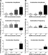
Both GYS (A, C, and E) and SGRN (B, D, and F) altered contraction parameters when perfused through whole heart preparations. Thresholds, indicated by changes significantly greater than 0, for changes in at least some contraction parameters, were as low as 10−11 M in both peptides. A and B: contraction amplitude increased with increasing peptide concentration for both peptides. C and D: contraction frequency showed small (<20%) but significant increases in both peptides at concentrations below 10−8 M; at 10−8 M, however, frequency decreased significantly in GYS and was unaltered by perfusion with SGRN. E and F: contraction duration was essentially unchanged at low peptide concentrations, showing only a slight (<10%) decrease in GYS at 10−10 and 10−11 M. In contrast, when peptides were perfused through the heart at 10−8 M, contraction duration increased somewhat. *Values significantly different from 0; one-sample t-test; GYS 10−11 n = 23; GYS 10−10 n = 21; GYS 10−9 n = 11; GYS 10−8 n = 14; SGRN 10−11 n = 10; SGRN 10−10 n = 13; SGRN 10−9 n = 14; SGRN 10−8 n = 9.
Fig. 3.
Fig. 3.
When superfused over the isolated cardiac ganglion (CG), GYS and SGRN exerted different modulatory effects on the motor neuronal bursting pattern. A and B: cycle frequency was unchanged by superfusion with GYS (A), but showed dose-dependent decreases in response to SGRN (B). C and D: both peptides elicited increases in motor neuron burst duration. C: these increases were significantly greater than 0 at concentrations ranging from 10−9 to 10−7 M in GYS; this effect appeared to saturate at 10−8 M, showing no further increase when the concentration was raised from 10−8 to 10−7 M. D: in SGRN, threshold was 10−8 M, and this effect did not appear to saturate over the concentrations tested. E and F: burst duty cycle increased in both peptides. E: since cycle frequency did not change significantly in GYS, the pattern of increased in duty cycle paralleled that of the increases in burst duration. F: in SGRN, increases in duty cycle were significantly greater than 0 at both 10−8 and 10−7 M, but these increases were modest (<50%) since the increase in burst duration was accompanied by an increase in cycle period. *Values significantly different from 0; one-sample t-test; GYS 10−9 n = 9; GYS 10−8 n = 13; GYS 10−7 n = 9; SGRN 10−9 n = 9; SGRN 10−8 n = 10; SGRN 10−7 n = 10.
Fig. 4.
Fig. 4.
The overall patterns of changes in burst duration and duty cycle recorded when the CG was still embedded in the heart, and therefore subject to feedback, were similar in the two peptides, although thresholds and the extent of the changes differed. A and B: burst duration did not change in either peptide at concentrations of 10−10 or 10−11 M, but it increased somewhat at 10−9 M, and substantially (>100%) at 10−8M GYS (A). B: there was no change in burst duration in SGRN except at the highest concentration tested, 10−8 M. C and D: burst duty cycle increased in both peptides, but threshold differed. C: duty cycle increased in a dose-dependent manner in GYS, with significant changes at all concentrations tested. D: in SGRN, duty cycle, like burst duration, did not change at 10−11 or 10−10 M. Although burst duration did not increase in 10−9 M SGRN, duty cycle nonetheless increased because contraction frequency increased. At 10−8 M, the increase in burst duration coupled with no significant change in cycle frequency resulted in an increase in duty cycle. *Values significantly different from 0; one-sample t-test; GYS 10−11 n = 23; GYS 10−10 n = 21; GYS 10−9 n = 11; GYS 10−8 n = 14; SGRN 10−11 n = 10; SGRN 10−10 n = 13; SGRN 10−9 n = 14; SGRN 10−8 n = 9.
Fig. 5.
Fig. 5.
Perfusion with either GYS (A) or SGRN (B) at 10−8 M elicited large increases in the amplitude of contractions that resulted from controlled and consistent motor nerve stimulation; amplitude gradually returned to control values when the peptide was washed off. The CG was removed from the heart, and an anterolateral nerves was stimulated with 200-ms-long bursts of pulses (each 0.5 ms in duration) repeated at 1 Hz. To prevent damage from repeated stimulation, bursts were delivered in bouts of 15 bursts, followed by 45 s of recovery time. The cardiac muscle showed significant facilitation within each group of 15 bursts (bottom), and amplitude increased until it stabilized for the last few contractions. Both the initial contraction in a group and the final, facilitated contraction were larger in peptide than in control saline.
Fig. 6.
Fig. 6.
Both GYS (A and C) and SGRN (B and D) elicited dose-dependent changes in contraction amplitude when applied to stimulated nerve-muscle preparations in which motor neuronal input was held constant. Bursts of stimuli 200 ms in length were delivered to the motor nerve at a frequency of 1 Hz in bouts of 15 bursts separated by 45 s of recovery time. A and B: graphed are the averages of the last two contractions in each bout of 15 bursts; at this point, the heart had reached a steady-state amplitude. The effects of the two peptides on this preparation were very similar, with thresholds of ∼10−10 M, and increases to over 100% when the peptides were perfused at 10−8 M. C and D: both peptides elicited increases in contraction amplitude in response to the first train in each bout. Increases were significant at concentrations of 10−10 M in both peptides, and increased strongly when peptide concentration was increased from 10−9 M to 10−8 M (P < 0.0001, Tukey multiple comparisons). *Values significantly different from 0; one-sample t-test; GYS 10−11 n = 18; GYS 10−10 n = 18; GYS 10−9 n = 17; GYS 10−8 n = 13; SGRN 10−11 n = 10; SGRN 10−10 n = 12; SGRN 10−9 n = 21; SGRN 10−8 n = 11. First contraction sample sizes GYS 10−11 n = 13; GYS 10−10 n = 11; GYS 10−9 n = 9; GYS 10−8 n = 7; SGRN 10−11 n = 9; SGRN 10−10 n = 11; SGRN 10−9 n = 18; SGRN 10−8 n = 8.
Fig. 7.
Fig. 7.
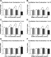
GYS and SGRN both altered facilitation of contraction amplitude recorded in response to repeated stimulation of the motor nerve (200-ms bursts delivered to the motor nerve at a frequency of 1 Hz in bouts of 15 bursts separated by 45 s of recovery time). Facilitation index was calculated as (amplitude of the later contraction)/(amplitude of the earlier contraction); thus an index or 1.0 indicates no facilitation. For each preparation, the facilitation index for the relevant contractions in control saline is depicted in the open bars; the facilitation index from the same preparations in peptide is shown in gray bars. A and B: facilitation over the course of the entire set of stimuli, i.e., from the first to the fifteenth contraction, was unaffected by low concentrations of either peptide, but decreased in the presence of both GYS (A) and SGRN (B) when the peptides were perfused through the heart at concentrations of 10−9 or 10−8 M. C and D: most of the decrease in facilitation in the presence of peptide occurred between the first and second contractions, with significant decreases in facilitation in the presence of GYS (C) at concentrations of 10−10 M and higher, and in the presence of SGRN (D) at concentrations of 10−9 and 10−8 M. E and F: to determine the extent to which GYS (E) and SGRN (F) affected facilitation after the second contraction, we calculated a facilitation index from the second to the fifteenth contraction. Neither peptide altered this facilitation index at any concentration. Open bars: control saline; gray bars, saline with peptide. Paired t-tests were used to compare each set of facilitation indices to their matched controls. *Significant differences; P < 0.05. GYS 10−11 n = 13; GYS 10−10 n = 13; GYS 10−9 n = 8; GYS 10−8 n = 5; SGRN 10−11 n = 9; SGRN 10−10 n = 11; SGRN 10−9 n = 17; SGRN 10−8 n = 7. Note that, although the facilitation index appears to increase in control saline in A and B, these differences between controls were not significant (ANOVA, P > 0.3).
Fig. 8.
Fig. 8.
Diagrammatic depiction of the cardiac neuromuscular system and its control and modulation by GYS and SGRN at high and low concentrations. Output from the CG acts on the cardiac muscle (open arrow) to elicit heart contractions. These contractions in turn are thought to control the generation of nitric oxide, which feeds back to the ganglion, inhibiting it to cause a decrease in cycle frequency (bottom arrow). Contractions also stretch the stretch-sensitive dendrites of the CG neurons; this is thought to result in positive feedback, and an increase in cycle frequency (top arrow). The effects of the peptides are shown in green (SGRN) and red (GYS) in each panel, with thicker arrows indicating effects that are predicted to be stronger. Tables list the observed effects of the peptides on the whole heart, the isolated CG, and the stimulated muscle preparation. A: in the presence of low (10−10 to 10−9 M) GYS, contraction amplitude increases peripherally; burst duration increases in the isolated CG, but not in the whole heart. Thus the effects on frequency appear to be mediated by an increase in the stretch feedback resulting from the peripheral enhancement of contraction amplitude. B: similar to A, in the presence of low (10−10 to 10−9 M) SGRN, contraction amplitude increases peripherally. Thus the effects on frequency appear to be mediated by an increase in the stretch feedback resulting from the peripheral enhancement of contraction amplitude. C: higher concentrations of GYS (10−8 M) elicit not only an increase in peripheral contraction amplitude, but also increases in burst duration within the CG itself, sufficient to result in increased burst duration in the whole heart. However, although there is no direct effect on cycle frequency in the CG, cycle frequency in the whole heart decreases, suggesting that the impact of the nitric oxide feedback pathway predominates (thick green arrow). D: higher concentrations of SGRN (10−8 M) elicit not only an increase in peripheral contraction amplitude, but, like GYS, increases in burst duration within the CG itself, which in turn leads to increased burst duration in the whole heart. In spite of the increased burst duration and the decreased cycle frequency in the isolated CG, cycle frequency remains unchanged in the whole heart, suggesting that the impact of the stretch feedback pathway predominates (thick red arrow). Because burst duration increases while frequency remains constant, duty cycle increases, which may also contribute to the increases in contraction amplitude. +, Increase; −, decrease; 0, no significant change.
Fig. 9.
Fig. 9.
Heat-map depiction of the neuromuscular transform for the heart of H. americanus, showing the general changes that are predicted in both duty cycle and cycle frequency in the two peptides, GYS (green) and SGRN (red). The neuromuscular transform heat-map illustrates the normalized contraction amplitude that resulted from stimulating the motor nerve over a range of cycle frequency and duty cycle pairs. Lighter colors represent larger contractions. The average starting values for activity in the whole heart are represented by circles, which are slightly offset so that they are visible. The average changes in each parameter are represented by the arrows. A: perfusion of either peptide at 10−9 M resulted in increases in both duty cycle and burst frequency. The increase in duty cycle would be predicted to cause an increase in contraction amplitude, while the increase in cycle frequency would be predicted to cause a decrease in amplitude. B: when the peptides were perfused at 10−8 M, GYS elicited an increase in duty cycle, but a decrease in frequency; SGRN elicited an increase in duty cycle, but did not significantly alter frequency. Globally, these changes are predicted to result in increased contraction amplitude, with a larger increase in GYS than in SGRN. [Neuromuscular transform heat-map modified from Williams et al. 2013 with permission.]

References

    1. Alexandrowicz JS. The innervation of the heart of the Crustacea. I Decapoda. Q J Microsc Sci 75: 181–249, 1932.
    1. Amendola J, Woodhouse A, Martin-Eauclaire MF, Goaillard JM. Ca(2)(+)/cAMP-sensitive covariation of I(A) and I(H) voltage dependences tunes rebound firing in dopaminergic neurons. J Neurosci 32: 2166–2181, 2012. - PMC - PubMed
    1. Anderson M, Cooke IM. Neural activation of the heart of the lobster Homarus americanus. J Exp Biol 55: 449–468, 1971. - PubMed
    1. Beilin SA, Pasztor VM. Modulation of a rhythmically active crayfish muscle by the neuropeptide proctolin. Can J Zool 67: 73–81, 1989.
    1. Bendena WG, Garside CS, Yu CG, Tobe SS. Allatostatins: diversity in structure and function of an insect neuropeptide family. Ann NY Acad Sci 814: 53–66, 1997. - PubMed

Publication types

LinkOut - more resources