. 2015 Jan 15;89(2):1329-39.
doi: 10.1128/JVI.01730-14. Epub 2014 Nov 12.

Distinct characteristics of endometrial and decidual macrophages and regulation of their permissivity to HIV-1 infection by SAMHD1

Affiliations

Distinct characteristics of endometrial and decidual macrophages and regulation of their permissivity to HIV-1 infection by SAMHD1

Héloïse Quillay et al. J Virol. .

Abstract

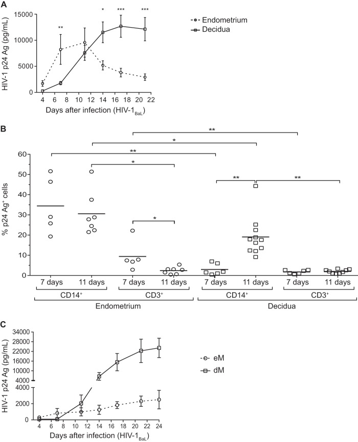
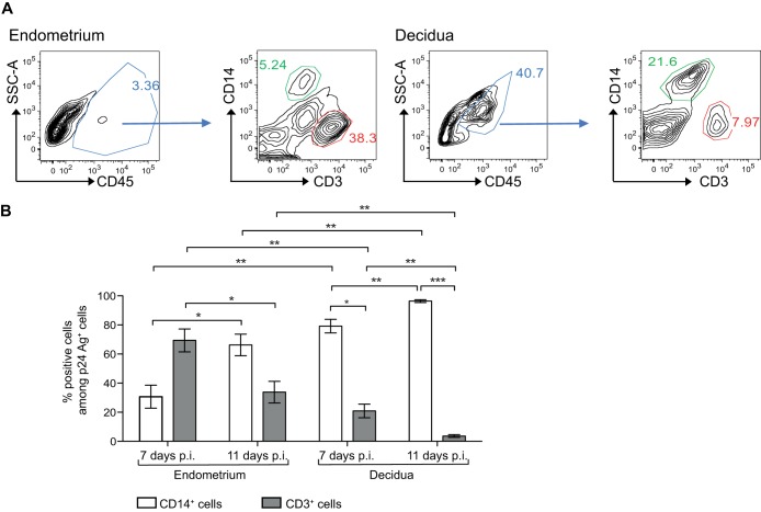
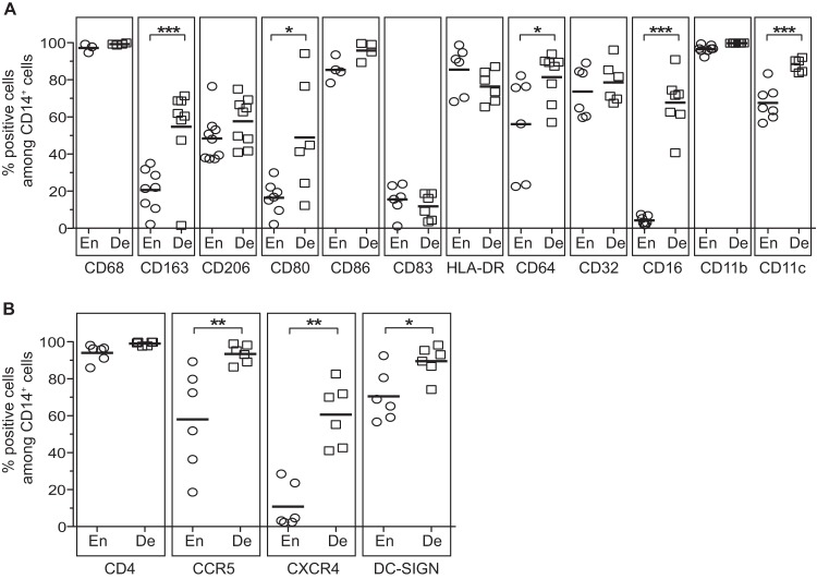
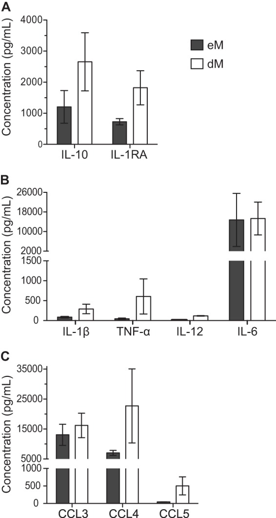
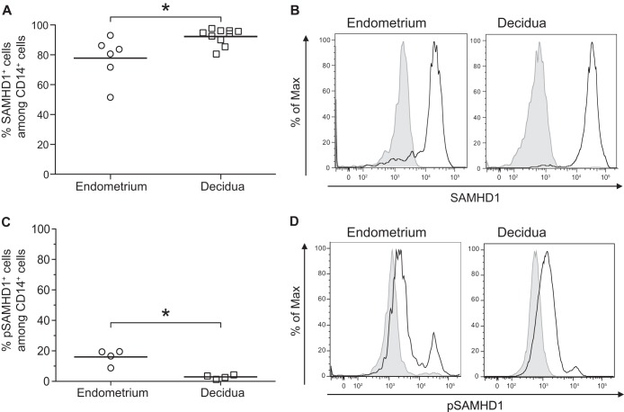
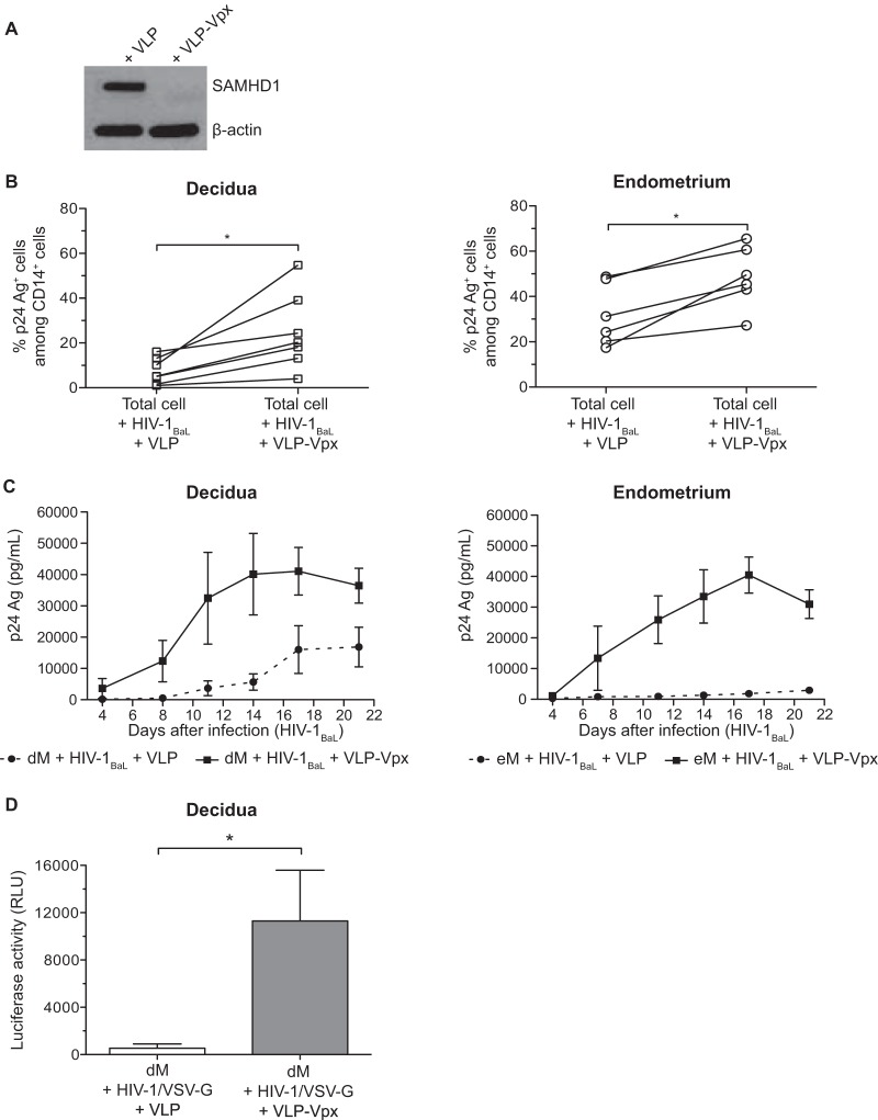
In order to develop strategies to prevent HIV-1 (human immunodeficiency virus type 1) transmission, it is crucial to better characterize HIV-1 target cells in the female reproductive tract (FRT) mucosae and to identify effective innate responses. Control of HIV-1 infection in the decidua (the uterine mucosa during pregnancy) can serve as a model to study natural mucosal protection. Macrophages are the main HIV-1 target cells in the decidua. Here we report that in vitro, macrophages and T cells are the main HIV-1 targets in the endometrium in nonpregnant women. As reported for decidual macrophages (dM), endometrial macrophages (eM) were found to have an M2-like phenotype (CD68+ CD163+ CD206+ IL-10high). However, eM and dM may belong to different subpopulations, as they differently express certain markers and secrete different amounts of proinflammatory and anti-inflammatory cytokines. We observed strong expression of the SAMHD1 restriction factor and weak expression of its inactive form (pSAMHD1, phosphorylated at residue Thr592) in both eM and dM. Infection of macrophages from both tissues was enhanced in the presence of the viral protein Vpx, suggesting a role for SAMHD1 in the restriction of HIV-1 infection. This study and further comparisons of the decidua with FRT mucosae in nonpregnant women should help to identify mechanisms of mucosal protection against HIV-1 infection.

Importance: The female reproductive tract mucosae are major portals of HIV-1 entry into the body. The decidua (uterine mucosa during pregnancy) can serve as a model for studying natural mucosal protection against HIV-1 transmission. A comparison of target cells and innate responses in the decidua versus the endometrium in nonpregnant women could help to identify protective mechanisms. Here, we report for the first time that macrophages are one of the main HIV-1 target cells in the endometrium and that infection of macrophages from both the endometrium and the decidua is restricted by SAMHD1. These findings might have implications for the development of vaccines to prevent HIV-1 mucosal transmission.


Figures

FIG 1
FIG 1
Infection of endometrial and decidual mononuclear cells and macrophages. (A and B) Endometrial and decidual mononuclear cells were exposed to HIV-1BaL at an MOI of 10−3. (A) The HIV-1 p24 Ag was measured by ELISA in the culture supernatants of endometrial (n = 11) and decidual (n = 16) mononuclear cells over time. The mean and the standard error of the mean of the viral production are displayed. (B) Intracellular staining of the p24 Ag was performed on days 7 and 11 postinfection in endometrial and decidual mononuclear cells. The percentage of p24 Ag+ cells among CD14+ cells or CD3+ cells is shown (n = 5 on day 7 and n = 7 on day 11 for the endometrium; n = 6 on day 7 and n = 11 on day 11 for the decidua). Each circle represents one endometrial sample and each square one decidual sample. The means are displayed. *, P < 0.05; **, P < 0.005; ***, P < 0.0005. (C) Endometrial (eM, n = 4) and decidual (dM, n = 17) purified macrophages were exposed to HIV-1BaL at an MOI of 10−3. The HIV-1 p24 Ag was measured by ELISA in the culture supernatants over time.
FIG 2
FIG 2
Percentage of CD14+ cells and CD3+ cells among endometrial and decidual HIV-1-infected cells. (A) Flow cytometry graphs show the gating strategy for the identification of CD14+ cells (green gate) and CD3+ cells (red gate). (B) Endometrial and decidual mononuclear cells were exposed to HIV-1BaL at an MOI of 10−3. Intracellular staining of the p24 Ag was performed on days 7 and 11 postinfection (p.i.). The mean percentages of CD14+ cells or CD3+ cells among p24 Ag+ cells are displayed (n = 5 on day 7 and n = 7 on day 11 for the endometrium; n = 6 on day 7 and n = 11 on day 11 for the decidua). Bars indicate standard errors of the means. *, P < 0.05; **, P < 0.005.
FIG 3
FIG 3
Phenotypes of endometrial and decidual CD14+ cells. Expression of antigen-presenting cell markers (A) and of HIV-1 receptor and coreceptors (B) was analyzed on CD45+ CD14+ cells by flow cytometry stainings of total endometrial or decidual cells. The mean percentages of expression of these markers among the CD45+ CD14+ cells from the endometrium (En) or the decidua (De) are depicted. Each circle represents one endometrial sample and each square one decidual sample. *, P < 0.05; **, P < 0.005; ***, P < 0.0005.
FIG 4
FIG 4
Cytokine and chemokine secretion by purified endometrial and decidual macrophages. Anti-inflammatory cytokine (A), proinflammatory cytokine (B), and chemokine (C) concentrations were analyzed by a Luminex assay in supernatants from purified eM and dM after 72 h of culture. The means of 3 samples and the standard errors of the means are depicted.
FIG 5
FIG 5
Expression of SAMHD1 in decidual and endometrial macrophages. The expression of SAMHD1 and the phosphorylated form of SAMHD1 at residue Thr592 (pSAMHD1) was analyzed on CD45+ CD14+ cells by flow cytometry staining of total endometrial or decidual cells. An isotype-matched IgG control was used to confirm the specificity of the anti-SAMHD1 and the anti-pSAMHD1 Thr592. The mean percentages of SAMHD1 (A) and pSAMHD1 (C) expression among the CD14+ cells are displayed. Each circle represents one endometrial sample (n = 6 for SAMHD1 and n = 4 for pSAMHD1) and each square one decidual sample (n = 10 for SAMHD1 and n = 4 for pSAMHD1). *, P < 0.05. (B and D) A representative histogram is shown for one endometrial sample and for one decidual sample. The black line represents SAMHD1 (B) or pSAMHD1 (D) expression and shaded gray the isotype-matched control IgG.
FIG 6
FIG 6
SAMHD1 degradation and infection of decidual and endometrial cells in the presence of VLP-Vpx. (A) Control VLP or VLP-Vpx was added to purified decidual CD14+ cells, and after 72 h, SAMHD1 expression was analyzed by Western blotting. (B) Decidual and endometrial cells were exposed to HIV-1BaL at an MOI of 10−3 in the presence of control VLP or VLP-Vpx. Intracellular staining of the p24 Ag was performed on day 11 postinfection. The percentage of p24 Ag+ cells among CD14+ cells of each sample (n = 6 for the endometrium and n = 7 for the decidua) is displayed. (C) Purified eM and dM were exposed to HIV-1BaL at an MOI of 10−3 in the presence of control VLP (dashed line) or VLP-Vpx (full line). Values represent the means of the p24 Ag concentrations over time (n = 3 for the endometrium and n = 4 for the decidua). (D) Purified dM were exposed to the HIV-1/VSV-G pseudotype (600 ng of p24 Ag/106 cells) in the presence of control VLP or VLP-Vpx. The mean luciferase activity is given for 6 samples, in relative light units (RLU). Bars indicate standard errors of the means. *, P < 0.05.

References

    1. UNAIDS. 2013. UNAIDS report on the global AIDS epidemic 2013. UNAIDS, Geneva, Switzerland.
    1. UNAIDS. 2010. Women, girls, gender equality and HIV. UNAIDS, Geneva, Switzerland.
    1. Li Q, Estes JD, Schlievert PM, Duan L, Brosnahan AJ, Southern PJ, Reilly CS, Peterson ML, Schultz-Darken N, Brunner KG, Nephew KR, Pambuccian S, Lifson JD, Carlis JV, Haase AT. 2009. Glycerol monolaurate prevents mucosal SIV transmission. Nature 458:1034–1038. doi:10.1038/nature07831. - DOI - PMC - PubMed
    1. Joag SV, Adany I, Li Z, Foresman L, Pinson DM, Wang C, Stephens EB, Raghavan R, Narayan O. 1997. Animal model of mucosally transmitted human immunodeficiency virus type 1 disease: intravaginal and oral deposition of simian/human immunodeficiency virus in macaques results in systemic infection, elimination of CD4+ T cells, and AIDS. J Virol 71:4016–4023. - PMC - PubMed
    1. Howell AL, Edkins RD, Rier SE, Yeaman GR, Stern JE, Fanger MW, Wira CR. 1997. Human immunodeficiency virus type 1 infection of cells and tissues from the upper and lower human female reproductive tract. J Virol 71:3498–3506. - PMC - PubMed

Publication types

Substances

LinkOut - more resources