. 2015 Jan 15;89(2):1428-38.
doi: 10.1128/JVI.02898-14. Epub 2014 Nov 12.

A cryo-electron microscopy study identifies the complete H16.V5 epitope and reveals global conformational changes initiated by binding of the neutralizing antibody fragment

Affiliations

A cryo-electron microscopy study identifies the complete H16.V5 epitope and reveals global conformational changes initiated by binding of the neutralizing antibody fragment

Hyunwook Lee et al. J Virol. .

Abstract

Human papillomavirus 16 (HPV16) is a worldwide health threat and an etiologic agent of cervical cancer. To understand the antigenic properties of HPV16, we pursued a structural study to elucidate HPV capsids and antibody interactions. The cryo-electron microscopy (cryo-EM) structures of a mature HPV16 particle and an altered capsid particle were solved individually and as complexes with fragment of antibody (Fab) from the neutralizing antibody H16.V5. Fitted crystal structures provided a pseudoatomic model of the virus-Fab complex, which identified a precise footprint of H16.V5, including previously unrecognized residues. The altered-capsid-Fab complex map showed that binding of the Fab induced significant conformational changes that were not seen in the altered-capsid structure alone. These changes included more ordered surface loops, consolidated so-called "invading-arm" structures, and tighter intercapsomeric connections at the capsid floor. The H16.V5 Fab preferentially bound hexavalent capsomers likely with a stabilizing effect that directly correlated with the number of bound Fabs. Additional cryo-EM reconstructions of the virus-Fab complex for different incubation times and structural analysis provide a model for a hyperstabilization of the capsomer by H16.V5 Fab and showed that the Fab distinguishes subtle differences between antigenic sites.

Importance: Our analysis of the cryo-EM reconstructions of the HPV16 capsids and virus-Fab complexes has identified the entire HPV.V5 conformational epitope and demonstrated a detailed neutralization mechanism of this clinically important monoclonal antibody against HPV16. The Fab bound and ordered the apical loops of HPV16. This conformational change was transmitted to the lower region of the capsomer, resulting in enhanced intercapsomeric interactions evidenced by the more ordered capsid floor and "invading-arm" structures. This study advances the understanding of the neutralization mechanism used by H16.V5.


Figures

FIG 1
FIG 1
Cryo-EM 3D reconstructions of HPV16 and virus-H16.V5 Fab complex. (A) The 3D reconstruction of mature virus had a 600-Å diameter. (B) The virus-Fab complex map shows the mature virus decorated by H16.V5 Fab molecules and reveals that the Fabs bound the pentavalent capsomers on the 5-fold vertex. Surface-rendered images are oriented on an icosahedral 5-fold symmetry axis, and internal DNA was computationally removed (middle) to visualize the protein capsid shell. The rendered surfaces were radially colored according to the key and are displayed at a contour level of 1.0 σ above background. Central sections in standard orientation (on the 2-fold axis, with symmetry axes indicated in black) show the quality of the density (right).
FIG 2
FIG 2
The pseudoatomic model was used to identify the Fab footprint. (A) The crystal structure of HPV16 pentamer (hexavalent and pentavalent capsomers are in green and yellow, respectively) and the atomic model of H16.V5 variable-domain (blue, heavy chain; red, light chain) were fitted into the density map. Fab-1 and -2 were replicated from the fitted Fab-3. The mature-virus–Fab cryo-EM density map is shown in gray mesh. (B) The side view of the Fab-1 and -2 bound on the pentavalent and hexavalent capsomers showed the steric clash between the two Fabs. The bound H16.V5 forms an angle of 47° respective to an axis through the center of the capsomer. (C) Each Fab binds across two L1 capsid proteins that are adjacent to one another. The contacting residues on the capsomeric surface are highlighted. Each L1 protein of the hexavalent capsomer is colored differently, and the pentavalent capsomer is in gray. The boundary of each Fab-binding residue is marked by a black dashed line. (D) The H16.V5 Fab bound 17 residues across five loops from two neighboring L1 proteins. The contacting residues are shown as spheres with the residue names and loops labeled.
FIG 3
FIG 3
Cryo-EM images of the altered capsids and the capsid-Fab complexes with their corresponding 3D maps. Representative areas of the cryomicrographs illustrate homogeneous, intact capsids that were used for image reconstructions (A and C) and altered capsids incubated with excess H16.V5 Fab molecules (B and D). The altered capsids showed the characteristic puffy, inflated capsomers of an immature VLP (18). H16.V5 Fabs occupied all 360 binding sites of the capsid, but the Fab densities on the pentavalent capsomers were weaker than the densities on hexavalent capsomers. The rendered surfaces were radially colored according to the key and displayed at a contour level of 1.0 σ above background.
FIG 4
FIG 4
Central sections show Fab-induced conformational changes to HPV. The three white circles indicate radii of 220, 260, and 300 Å on the central sections of the 3D maps of the altered capsid (A) and corresponding capsid-Fab complex (B). Significant differences were visualized within the 260- to 300-Å radii. Specifically, the HPV16 capsid had smeared density, whereas within the same radii of the complex map there were sharp, distinct features, indicating that surface loop structures had become more ordered upon Fab binding. Icosahedral symmetry axes are indicated in the central section, and the symmetry bars represent 50 nm.
FIG 5
FIG 5
The conformational change induced by Fab binding is illustrated by the alteration of densities and is more significant in the altered capsid than in mature virus. For the top panels, the reconstructions of the altered capsid (A), the capsid-Fab complex (B), the mature virus (C), and the virus-Fab complex (D) were radially projected at 280 Å. The pentavalent capsomers are indicated by yellow arrows. In altered capsids, Fab binding induced conformational changes on the hexavalent capsomers, whereas the change was attenuated on the pentavalent capsomers (A and B). The Fab-induced conformational changes were more significant in the altered capsid than in the mature virus (C and D). The bottom panels represent stereographic projections at the same radii on which the polar angles θ and ϕ represent latitude and longitude, respectively. The icosahedral asymmetric unit of the virus is indicated by the black triangular boundary, and the projections are colored according to the electron density height in the corresponding color keys.
FIG 6
FIG 6
Fab-induced stabilization of the capsid was shown by enhanced intercapsomeric connections. The reconstructions of the altered capsid (A), the capsid-Fab complex (B), the mature virus (C), and the virus-Fab complex (D) were radially projected at 265 Å (top), 257 Å (middle), and 240 Å (bottom) to show the invading arms and the capsid floor regions of the capsids. The invading-arm structures were significantly enhanced, and the electron densities at the icosahedral 3-fold axes of the capsid floor (yellow circles) were rearranged after Fab binding in altered capsids. The standard orientations of the capsids are the same as in Fig. 5.
FIG 7
FIG 7
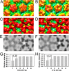
Fab preference for hexavalent capsomers was more pronounced in the altered HPV capsids. The pentavalent and hexavalent capsomers (outlined in black) of the 3D reconstructions of altered (A) and mature (B) virus showed marked density differences. (C and D) The capsid-Fab complexes had different Fab densities on the pentavalent and hexavalent capsomers, a difference that was more pronounced for the altered-capsid–Fab complex. The rendered surfaces were radially colored according to the color key in Fig. 1. (E and F) Radial projections of the capsid-Fab complex maps at the 310-Å radii show that Fabs bound on pentavalent capsomers have weaker intensities than those bound on hexavalent capsomers. (G and H) The relative mean intensities of the six Fabs bound on an icosahedral asymmetric unit of the capsid were measured and normalized by the mean intensity of Fab-4 (numbers are shown in panel E). The relative intensities of the Fab-1 and -2 quantify the preference of Fab for the binding sites on altered capsid (G) and mature virus (H).
FIG 8
FIG 8
Longer Fab incubation with virus enhanced the Fab binding to the hexavalent capsomer. Cryo-EM densities for the Fab-1 (left) and Fab-2 (right) on the pentavalent and hexavalent capsomers are shown for altered-virus–Fab (A) and mature-virus–Fab (B to D) complexes. The mature virus was incubated for 5 min (B), 1 h (C), and 72 h (D). The relative intensities of Fab-1 and -2 (numbers in each panel) indicate that as the incubation time increased, the Fab binding to the hexavalent capsomers increased. The rendered surfaces were radially colored according to the color key in Fig. 1 and presented at a contour level of 1.0 σ above background.

References

    1. Walboomers JM, Jacobs MV, Manos MM, Bosch FX, Kummer JA, Shah KV, Snijders PJ, Peto J, Meijer CJ, Muñoz N. 1999. Human papillomavirus is a necessary cause of invasive cervical cancer worldwide. J Pathol 189:12–19. doi:10.1002/(SICI)1096-9896(199909)189:1<12::AID-PATH431>3.0.CO;2-F. - DOI - PubMed
    1. Woodman CBJ, Collins SI, Young LS. 2007. The natural history of cervical HPV infection: unresolved issues. Nat Rev Cancer 7:11–22. doi:10.1038/nrc2050. - DOI - PubMed
    1. zur Hausen H. 2009. Papillomaviruses in the causation of human cancers—a brief historical account. Virology 384:260–265. doi:10.1016/j.virol.2008.11.046. - DOI - PubMed
    1. Lorincz AT, Reid R, Jenson AB, Greenberg MD, Lancaster W, Kurman RJ. 1992. Human papillomavirus infection of the cervix: relative risk associations of 15 common anogenital types. Obstet Gynecol 79:328–337. doi:10.1097/00006250-199203000-00002. - DOI - PubMed
    1. Bosch FX, Manos MM, Muñoz N, Sherman M, Jansen AM, Peto J, Schiffman MH, Moreno V, Kurman R, Shan KV. 1995. Prevalence of human papillomavirus in cervical cancer: a worldwide perspective. J Natl Cancer Inst 87:796–802. doi:10.1093/jnci/87.11.796. - DOI - PubMed

Publication types