. 2015 Feb;89(3):1550-63.
doi: 10.1128/JVI.02949-14. Epub 2014 Nov 12.

Biological and protective properties of immune sera directed to the influenza virus neuraminidase

Affiliations

Biological and protective properties of immune sera directed to the influenza virus neuraminidase

Stefan J Halbherr et al. J Virol. 2015 Feb.

Abstract

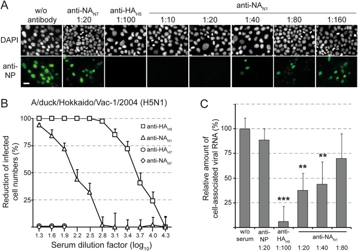
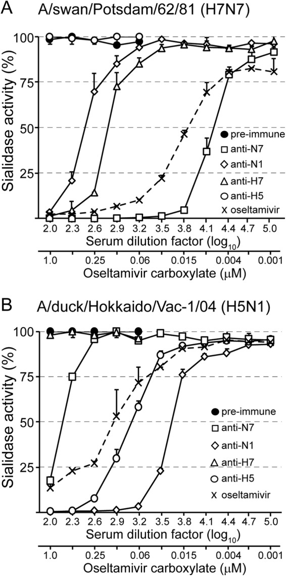
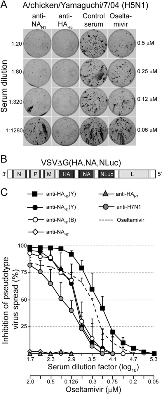
The envelope of influenza A viruses contains two large antigens, hemagglutinin (HA) and neuraminidase (NA). Conventional influenza virus vaccines induce neutralizing antibodies that are predominantly directed to the HA globular head, a domain that is subject to extensive antigenic drift. Antibodies directed to NA are induced at much lower levels, probably as a consequence of the immunodominance of the HA antigen. Although antibodies to NA may affect virus release by inhibiting the sialidase function of the glycoprotein, the antigen has been largely neglected in past vaccine design. In this study, we characterized the protective properties of monospecific immune sera that were generated by vaccination with recombinant RNA replicon particles encoding NA. These immune sera inhibited hemagglutination in an NA subtype-specific and HA subtype-independent manner and interfered with infection of MDCK cells. In addition, they inhibited the sialidase activities of various influenza viruses of the same and even different NA subtypes. With this, the anti-NA immune sera inhibited the spread of H5N1 highly pathogenic avian influenza virus and HA/NA-pseudotyped viruses in MDCK cells in a concentration-dependent manner. When chickens were immunized with NA recombinant replicon particles and subsequently infected with low-pathogenic avian influenza virus, inflammatory serum markers were significantly reduced and virus shedding was limited or eliminated. These findings suggest that NA antibodies can inhibit virus dissemination by interfering with both virus attachment and egress. Our results underline the potential of high-quality NA antibodies for controlling influenza virus replication and place emphasis on NA as a vaccine antigen.

Importance: The neuraminidase of influenza A viruses is a sialidase that acts as a receptor-destroying enzyme facilitating the release of progeny virus from infected cells. Here, we demonstrate that monospecific anti-NA immune sera inhibited not only sialidase activity, but also influenza virus hemagglutination and infection of MDCK cells, suggesting that NA antibodies can interfere with virus attachment. Inhibition of both processes, virus release and virus binding, may explain why NA antibodies efficiently blocked virus dissemination in vitro and in vivo. Anti-NA immune sera showed broader reactivity than anti-HA sera in hemagglutination inhibition tests and demonstrated cross-subtype activity in sialidase inhibition tests. These remarkable features of NA antibodies highlight the importance of the NA antigen for the development of next-generation influenza virus vaccines.


Figures

FIG 1
FIG 1
Expression of functionally active NA antigen using propagation-incompetent virus replicon particles. (A) Genome maps of recombinant VSV vectors. VSV*ΔG contains five transcription units encoding the nucleoprotein N, the phosphoprotein P, the matrix protein M, eGFP, and the large RNA polymerase protein L. Note that VSV*ΔG lacks the gene encoding the VSV envelope glycoprotein G. VSV*ΔG(NA) expresses influenza virus NA antigen from the fourth gene position, while GFP is expressed from an additional transcription unit downstream of NA. (B) Immunofluorescence analysis of Vero cells infected with either VSV*ΔG, VSV*ΔG(NAN1), or VSV*ΔG(NAN7). At 6 h p.i., the cells were incubated at 4°C with the indicated immune sera, washed with PBS, and fixed with formalin. Antibodies bound to cell surface NA were visualized with anti-chicken IgY serum conjugated with Alexa Fluor 546 (red fluorescence). Expression of GFP is indicated by green fluorescence. The scale bar represents 24 μm. (C) At 12 h p.i. with the indicated replicon particles, Vero cells were incubated with the sialidase substrate MU-NANA for 20 min at 37°C. Fluorescence was recorded at 355 nm for excitation and 460 nm for emission. The asterisk denotes significantly different sialidase activities (P < 0.05); ns, nonsignificantly different. The error bars indicate standard deviations. (D) Quantification of serum antibody levels. SPF chickens (n = 4/5) were immunized with either inactivated or live A/duck/Hokkaido/Vac-1/04 (H5N1) via the indicated vaccination routes. In addition, chickens were immunized i.m. with VRPs expressing either the NA antigen of A/chicken/Yamaguchi/7/04 (H5N1), the HA antigen of A/duck/Hokkaido/Vac-1/04 (H5N1), or GFP as a control (VSV*ΔG). With the exception of animals receiving live virus via the intratracheal route, all chickens were immunized a second time 3 weeks after the first immunization. Immune sera were collected 3 weeks after the boost and titrated using competitive N1 and H5 ELISAs. The scatter plots show individual and mean antibody titers. The asterisk indicates significantly different values (P < 0.05).
FIG 2
FIG 2
Anti-NA antibodies interfere with influenza virus infection. (A) A/chicken/Yamaguchi/7/04 (H5N1) was incubated with MDCK cell monolayers (0.5 TCID50/cell) for 1 h at 4°C in the presence or absence of the indicated immune sera. The cells were washed, incubated for 5 h at 37°C, fixed with formalin, and permeabilized with Triton X-100. The influenza virus NP antigen was detected by indirect immunofluorescence. The nuclei were stained with DAPI. The scale bar indicates 20 μm. (B) MDCK cells were infected with A/duck/Hokkaido/Vac-1/04 (H5N1) (MOI = 0.0025) in the presence of immune serum directed to HAH5 or NAN1 of A/chicken/Yamaguchi/7/04 (H5N1). Immune sera directed to HAH7 of A/turkey/Italy/3675/99 (H7N1) and NAN7 of A/swan/Potsdam/62/81 (H7N7) were used as controls. The cells were washed, maintained for 24 h in the absence of antibodies, and fixed. Infected cells were visualized by immunostaining with a monoclonal antibody directed to the influenza virus NP antigen. The number of infected cells was calculated as a percentage of the cells infected in the presence of nonimmune serum. The plaque reduction test was run in quadruplicate. Mean values and standard deviations are shown. (C) A/chicken/Yamaguchi/7/04 (H5N1) was incubated with MDCK cell monolayers (0.0025 TCID50/cell) for 1 h at 4°C in the presence or absence of the indicated immune sera. The cells were washed, total RNA was extracted, and viral RNA segment 7 was detected by quantitative RT-PCR. Virus bound to cells in the absence of immune serum was used as a reference (100% virus binding). The mean values and standard deviations of three independent experiments, each run in duplicate, are shown. The asterisks denote significantly different threshold cycle (CT) values compared to virus incubation in the absence of serum (**, P < 0.005; ***, P < 0.0005).
FIG 3
FIG 3
Anti-NA antibodies inhibit the sialidase of influenza virions. (A) A/swan/Potsdam/62/81 (H7N7) was mixed with serially diluted preimmune serum or immune serum raised against either NAN7 of A/swan/Potsdam/62/81 (H7N7), NAN1 of A/chicken/Yamaguchi/7/04 (H5N1), HAH7 of A/turkey/Italy/3675/99 (H7N1), or HAH5 of A/chicken/Yamaguchi/7/04 (H5N1). In parallel, virus was mixed with oseltamivir carboxylate at the indicated concentrations. The mixtures were incubated for 18 h at 37°C with immobilized glycophorin A. Desialylation of glycophorin A was detected by subsequent incubations with biotin-labeled peanut agglutinin, streptavidin-peroxidase conjugate, and TMB peroxidase substrate. (B) A/duck/Hokkaido/Vac-1/04 (H5N1) was mixed with the indicated immune sera or oseltamivir carboxylate prior to incubation with immobilized glycophorin A. Experiments were performed twice, each time with serum quadruplicates. The mean values and standard deviations of a representative experiment are shown.
FIG 4
FIG 4
Anti-NA antibodies cross-react with NAN1s of various influenza viruses. Immune sera raised against the NA of either A/chicken/Yamaguchi/7/04 (H5N1) (A) or A/swine/Belzig/2/01 (H1N1) (B) were tested for the ability to inhibit the sialidases of the indicated viruses using immobilized glycophorin A as the substrate. The mean values and standard deviations of quadruplicate measurements are shown. The test was performed at least two times, and a representative experiment is depicted.
FIG 5
FIG 5
Anti-NA antibodies inhibit virus spread in vitro. (A) MDCK monolayers were infected with the highly pathogenic avian influenza virus A/chicken/Yamaguchi/7/04 (H5N1) (MOI = 0.0025). The cells were washed and maintained for 18 h at 37°C in the presence of either anti-NAN1, anti-HAH5, nonimmune serum, or oseltamivir carboxylate. The serum dilutions and oseltamivir carboxylate concentrations used are indicated on the left and right sides of the graph, respectively. The cells were fixed, permeabilized, and stained using a monoclonal antibody directed to influenza virus NP antigen and a peroxidase-labeled anti-mouse IgG serum. (B) Genome map of recombinant VSV pseudotype vector expressing HA and NA antigens of A/chicken/Yamaguchi/7/04 (H5N1) and a secreted luciferase (NLuc) from O. gracilirostris. The vector lacks the VSV glycoprotein G gene. (C) Inhibition of pseudotype virus spread. MDCK cells were infected with the propagation-competent HA/NA pseudotype virus (MOI = 0.005) and incubated in the presence of the indicated concentrations of either immune sera or oseltamivir carboxylate. At 48 h p.i., the luciferase activity released into the cell culture supernatant was determined. The luciferase expression relative to that of cells that were incubated without immune serum is shown. Mean values and standard deviations of three infection experiments are shown.
FIG 6
FIG 6
Vaccination with NA-recombinant VRP reduces virus shedding from infected chickens and inflammation. SPF chickens (n = 6/group) were immunized (i.m.) twice with either VSV*ΔG or VSV*ΔG(NAN7) using 108 FFU per dose. Three weeks after the second immunization, the chickens were infected via the intratracheal route with 107 TCID50 of A/swan/Potsdam/62/81 (H7N7). (A) Detection of α1-AGP 2 days after infection. Box-and-whisker plots indicate maximum, minimum, and median values. The asterisk denotes significantly different α1-AGP levels between the two animal groups. (B and C) Detection of challenge virus in oropharyngeal- and cloacal-swab samples collected from chickens vaccinated with VSV*ΔG (B) or VSV*ΔG(NAN7) (C). Swab samples were taken from the animals at daily intervals for a period of 7 days. RNA was extracted from the samples and analyzed for the presence of viral RNA segment 7 by quantitative RT-PCR. Maximum 45 threshold cycles minus the threshold cycles detected are shown (45-Ct). Mean values and standard deviations are indicated. The asterisk denotes significantly different viral RNA loads in swabs taken from VSV*ΔG(NAN7)-vaccinated animals compared to control animals.
FIG 7
FIG 7
Phylogenetic analysis of influenza virus N1 neuraminidases by ClustalW alignment (78) and subsequent neighbor-joining tree construction by MEGA 4 software (79). The GenBank accession numbers for the NA protein sequences used are as follows: BAD89307 for A/chicken/Yamaguchi/7/04 (H5N1), ACS93996 for A/duck/Italy/1447/05 (H1N1), ACZ45212 for A/turkey/Italy/4580/99 (H7N1), BAE94701 for A/duck/Hokkaido/Vac-1/04 (H5N1), ACQ63272 for A/California/07/09 (H1N1), CAA36475 for A/chicken/Rostock/8/34 (H7N1), AAF77036 for A/Brevig Mission/1/18 (H1N1), ACV52104 for A/swine/USA/1976/31 (H1N1), ACF41881 for A/New Caledonia/20/99 (H1N1), and NP_040981 for A/Puerto Rico/8/34 (H1N1). The Global Initiative on Sharing All Influenza Data (GISAID) accession number for the NA sequence of A/swine/Belzig/2/01 (H1N1) is EPI302494.The scale bar indicates the number of nucleotide substitutions per site.

References

    1. Krammer F, Palese P. 2013. Influenza virus hemagglutinin stalk-based antibodies and vaccines. Curr Opin Virol 3:521–530. doi:10.1016/j.coviro.2013.07.007. - DOI - PMC - PubMed
    1. Lotan R, Skutelsky E, Danon D, Sharon N. 1975. The purification, composition, and specificity of the anti-T lectin from peanut (Arachis hypogaea). J Biol Chem 250:8518–8523. - PubMed
    1. Xu X, Cox NJ, Bender CA, Regnery HL, Shaw MW. 1996. Genetic variation in neuraminidase genes of influenza A (H3N2) viruses. Virology 224:175–183. doi:10.1006/viro.1996.0519. - DOI - PubMed
    1. Luther P, Bergmann KC, Oxford JS. 1984. An investigation of antigenic drift of neuraminidases of influenza A (H1N1) viruses. J Hyg 92:223–229. doi:10.1017/S002217240006424X. - DOI - PMC - PubMed
    1. Kilbourne ED, Johansson BE, Grajower B. 1990. Independent and disparate evolution in nature of influenza A virus hemagglutinin and neuraminidase glycoproteins. Proc Natl Acad Sci U S A 87:786–790. doi:10.1073/pnas.87.2.786. - DOI - PMC - PubMed

Publication types