. 2014 Nov 13;9(11):e112142.
doi: 10.1371/journal.pone.0112142. eCollection 2014.

Identification of a common non-apoptotic cell death mechanism in hereditary retinal degeneration

Affiliations

Identification of a common non-apoptotic cell death mechanism in hereditary retinal degeneration

Blanca Arango-Gonzalez et al. PLoS One. .

Abstract

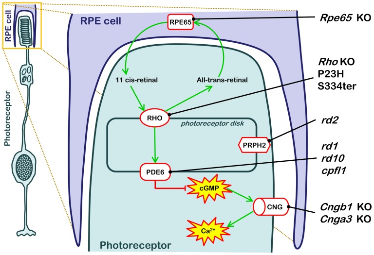
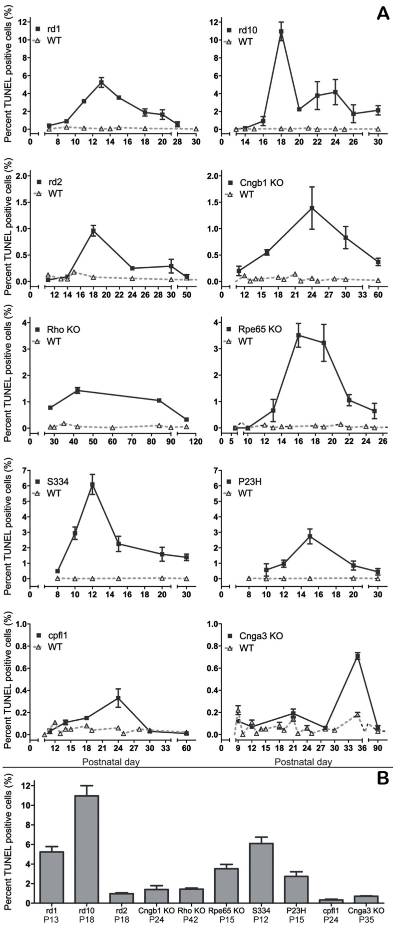
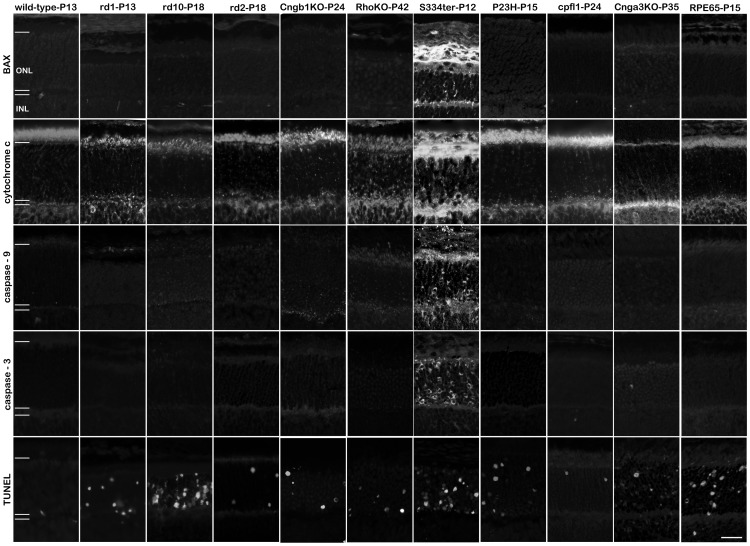
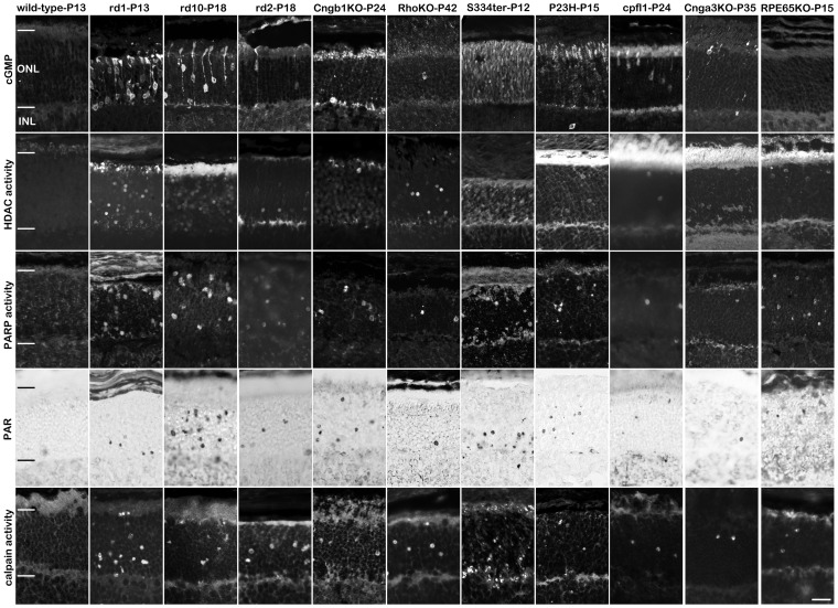
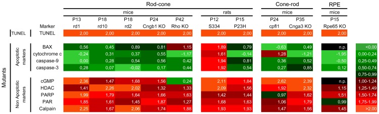
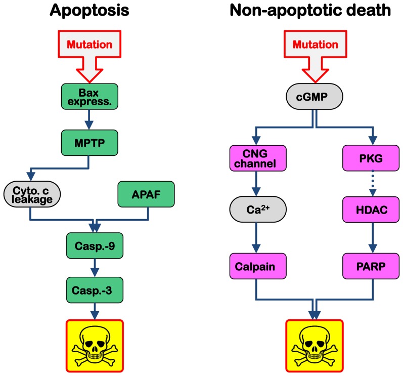
Cell death in neurodegenerative diseases is often thought to be governed by apoptosis; however, an increasing body of evidence suggests the involvement of alternative cell death mechanisms in neuronal degeneration. We studied retinal neurodegeneration using 10 different animal models, covering all major groups of hereditary human blindness (rd1, rd2, rd10, Cngb1 KO, Rho KO, S334ter, P23H, Cnga3 KO, cpfl1, Rpe65 KO), by investigating metabolic processes relevant for different forms of cell death. We show that apoptosis plays only a minor role in the inherited forms of retinal neurodegeneration studied, where instead, a non-apoptotic degenerative mechanism common to all mutants is of major importance. Hallmark features of this pathway are activation of histone deacetylase, poly-ADP-ribose-polymerase, and calpain, as well as accumulation of cyclic guanosine monophosphate and poly-ADP-ribose. Our work thus demonstrates the prevalence of alternative cell death mechanisms in inherited retinal degeneration and provides a rational basis for the design of mutation-independent treatments.


Conflict of interest statement

Competing Interests: The authors declare that they have no conflict of interest.

Figures

Figure 1
Figure 1. RD animal models used and their genetic defects.
The cartoon illustrates the anatomical localization and metabolic consequences of the causative genetic mutations in the ten different RD animal models used in this study. RD causing mutations in these animal models interfere with the various stages of the phototransduction cascade, from the 11-cis-retinal recycling enzyme RPE65 (Rpe65 KO), via the light-sensitive Rhodopsin (Rho KO, P23H, S334ter), cGMP-hydrolyzing phosphodiesterase-6 (PDE6; rd1, rd10, cpfl1), the structural protein Peripherin (Prph2; rd2), to the cyclic-nucleotide-gated (CNG; Cngb1 KO, Cnga3 KO) channel that allows for Ca2+-influx.
Figure 2
Figure 2. Progression of cell death in inherited RD models.
Depending on the causative genetic insult, the temporal development of retinal degeneration is highly variable in the different animal models. The quantification of dying, TUNEL-positive photoreceptor cells in the outer nuclear layer (ONL) allowed determination of the evolution and the peak of photoreceptor death for each of these animal models (A). The peak was taken as reference point for the ensuing analysis of cell death mechanisms. The bar graph (B) shows a comparison of maximum peak heights for all ten RD models studied. Note the different scales in line graphs. Values are mean ± SEM from at least three different animals. See also Table S1 and S2.
Figure 3
Figure 3. Apoptosis in the retina is restricted to the S334ter rat model.
The analysis of BAX expression, mitochondrial cytochrome c release, activation of caspase-9 and -3 shows essentially no positive detection in 9 out of 10 animal models for hereditary retinal degeneration. The notable exception was the S334ter transgenic rat which harbours a mutation in the rhodopsin gene leading to a truncated protein and in which many photoreceptors were positive for apoptosis. In all other animal models, while there were cells displaying clear evidence for apoptosis, their numbers were within the wild-type levels, indicating that this was related to physiological, developmental cell death, which is characteristic for the postnatal rodent retina. Importantly, the numbers of apoptotic cells did not match the numbers of mutation-induced dying cells as evidenced by the TUNEL assay. Scale bar 20 µm.
Figure 4
Figure 4. Cell death in hereditary retinal degeneration is predominantly non-apoptotic.
In 10 out of 10 animal models for hereditary retinal degeneration, large numbers of photoreceptors display cGMP accumulation, HDAC and PARP activity, PAR accumulation, and calpain activity, respectively. Intriguingly, these non-apoptotic markers are prominent even in the S334ter retina, concomitant with this also showing signs of apoptosis. This suggests that in S334ter retina two different cell death mechanisms may run in parallel while in all other studied RD models the mutation-induced cell death followed a non-apoptotic mechanism. Scale bar 20 µm.
Figure 5
Figure 5. Heat map representing metabolic activities in different RD models.
The RD models were grouped according to the peak of degeneration, the cell type affected by the mutation (rod, cone, RPE), and species (mouse, rat). The number of TUNEL-positive cells in each model was normalized to 100, expressed as logarithm, and compared with the number of positively labelled cells for each marker. The heat map clearly illustrates the prevalence of non-apoptotic vs. apoptotic cell death in 9 out of 10 RD models. The S334ter rhodopsin mutant was unique, showing concurrent activation of both cell death pathways. n.p.: null positive. See also Table S1 and S2.
Figure 6
Figure 6. Two routes to cell death.
Classical apoptosis, such as it occurs in S334ter transgenic photoreceptors, involves a mutation-induced up-regulation and translocation of BAX protein to form the mitochondrial permeability transition pore (MPTP). This leads to leakage of cytochrome c from the mitochondria to the cytoplasm, where it combines with apoptotic protease activating factor (APAF) and caspase-9 to form the apoptosome, which in turn activates down-stream executioner caspases, including caspase-3. In 9/10 RD animal models investigated here, photoreceptor death followed a different route: mutation-induced up-regulation of cGMP on the one hand causes activation of the CNG channel, leading to Ca2+ influx and calpain activation. On the other hand cGMP-dependent activation of protein kinase G (PKG) is associated with histone deacetylase (HDAC) and poly-ADP-ribose-polymerase (PARP) activation. Importantly, this alternative, non-apoptotic cell death mechanism offers a number of novel targets for neuroprotection of photoreceptors.

References

    1. Orrenius S, Zhivotovsky B, Nicotera P (2003) Regulation of cell death: the calcium-apoptosis link. Nat Rev Mol Cell Biol 4: 552–565. - PubMed
    1. Chang GQ, Hao Y, Wong F (1993) Apoptosis: final common pathway of photoreceptor death in rd, rds, and rhodopsin mutant mice. Neuron 11: 595–605. - PubMed
    1. Marigo V (2007) Programmed cell death in retinal degeneration: targeting apoptosis in photoreceptors as potential therapy for retinal degeneration. Cell Cycle 6: 652–655. - PubMed
    1. Hamann S, Schorderet DF, Cottet S (2009) Bax-induced apoptosis in Leber's congenital amaurosis: a dual role in rod and cone degeneration. PLoS One 4: e6616 10.1371/journal.pone.0006616 - DOI - PMC - PubMed
    1. Yoshizawa K, Kiuchi K, Nambu H, Yang J, Senzaki H, et al. (2002) Caspase-3 inhibitor transiently delays inherited retinal degeneration in C3H mice carrying the rd gene. Graefes Arch Clin Exp Ophthalmol 240: 214–219. - PubMed

Publication types