. 2014 Nov 13;9(11):e113110.
doi: 10.1371/journal.pone.0113110. eCollection 2014.

Normal and functional TP53 in genetically stable myxoid/round cell liposarcoma

Affiliations

Normal and functional TP53 in genetically stable myxoid/round cell liposarcoma

Anders Ståhlberg et al. PLoS One. .

Abstract

Myxoid/round-cell liposarcoma (MLS/RCLS) is characterized by either the fusion gene FUS-DDIT3 or the less commonly occurring EWSR1-DDIT3 and most cases carry few or no additional cytogenetic changes. There are conflicting reports concerning the status and role of TP53 in MLS/RCLS. Here we analysed four MLS/RCLS derived cell lines for TP53 mutations, expression and function. Three SV40 transformed cell lines expressed normal TP53 proteins. Irradiation caused normal posttranslational modifications of TP53 and induced P21 expression in two of these cell lines. Transfection experiments showed that the FUS-DDIT3 fusion protein had no effects on irradiation induced TP53 responses. Ion Torrent AmpliSeq screening, using the Cancer Hotspot panel, showed no dysfunctional or disease associated alleles/mutations. In conclusion, our results suggest that most MLS/RCLS cases carry functional TP53 genes and this is consistent with the low numbers of secondary mutations observed in this tumor entity.


Conflict of interest statement

Competing Interests: AS declares stock ownership in TATAA Biocenter. CYL has been employed by Life Technologies and has patent applications pending at Thermo Fisher Scientific. DR has been employed by Life Technologies and is currently employed by Fluidigm, patent applications pending at both Thermo Fisher Scientific and Fluidigm, stock ownership in Fluidigm. SMC is employed by Thermo Fisher Scientific and has patent applications pending at Thermo Fisher Scientific. The authors confirm that these competing interests do not alter their adherence to PLOS ONE policies on sharing data and materials.

Figures

Figure 1
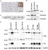
Figure 1. TP53 expression in MLS/RCLS.
(A) Immunohistochemistry analysis of TP53 expression in a representative MLS/RCLS case. Inset shows TP53 mutated endometrial carcinoma as positive control. Brown precipitate indicates TP53 expression. Scale bar shows 100 µm. (B) Western blot analysis of TP53 in MLS and HT1080 cell lines. Three different antibodies directed against the core (central transcription factor part), N- and C-terminal parts were used. Two distinct bands (53 and 56 kD, respectively) were detected for the core part of TP53, while only the shorter 53 kD band was detected using antibodies against N- and C-terminal part of TP53. (C) Schematic map of eleven exons in TP53. The translated region is shown as dotted line, and only the translated part of exon eleven is shown. Transcripts detected by reverse transcription PCR and sequencing are shown as black lines. Protein fragment analyses by mass spectroscopy for MLS 402-91 are shown as grey lines. (D) Irradiation effect on TP53 and P21 (CDKN1A) expression in four MLS/RCLS cell lines, wild type HT1080 cells and HT1080 cells expressing FUS-DDIT3 (HT1080 FUS-DDIT3). Antibody against core TP53 part was used. The 68 KD band corresponds to post-translationally modified TP53. MLS 402-91 and 1765-92 carry simian virus 40 large T antigen, while MLS 2645-94 was established using the complete SV40 virus. GAPDH is used as loading control, +/− indicate irradiated and control cell samples, respectively. Positions and sizes of reference proteins are indicated.
Figure 2
Figure 2. Immunofluorescence analysis of TP53 and P21 in irradiated and control cultured cells.
Percentage of (A) TP53 and (B) P21 positive cells are shown. Mean ± SEM of three experiments are shown. * and ** indicate 95% and 99% significance using student's t-test. The number of strongly TP53 stained cells was assessed for one experiment, marked with dot (•) in respective bar. Representative immunofluorescence images for MLS 402-91 are shown. Scale bar shows 50 µm. (C) Western blot analysis of FUS-DDIT3 in MLS cell lines are shown. Different sizes correspond to Type I (MLS 402-91 and DL 221), II (MLS 2645-94) and VI (MLS 1765-92) fusion proteins. GAPDH is used as internal control between samples when calculating the relative FUS-DDIT3 expression level. The lowest FUS-DDIT3 expression value (DL 221) was arbitrarily set to a value of one.

References

    1. Mertens F, Panagopoulos I, Mandahl N (2010) Genomic characteristics of soft tissue sarcomas. Virchows Arch 456: 129–139. - PubMed
    1. Aman P (1999) Fusion genes in solid tumors. Semin Cancer Biol 9: 303–318. - PubMed
    1. Åman P, Ron D, Mandahl N, Fioretos T, Heim S, et al. (1992) Rearrangement of the transcription factor gene CHOP in myxoid liposarcomas with t(12;16)(q13;p11). Genes Chromosom Cancer 5: 278–285. - PubMed
    1. Panagopoulos I, Höglund M, Mertens F, Mandahl N, Mitelman F, et al. (1996) Fusion of the EWS and CHOP genes in myxoid liposarcoma. Oncogene 12: 489–494. - PubMed
    1. Engström K, Willén H, Kåbjörn-Gustafsson C, Andersson C, Olsson M, et al. (2006) The myxoid/round cell liposarcoma fusion oncogene FUS-DDIT3 and the normal DDIT3 induce a liposarcoma phenotype in transfected human fibrosarcoma cells. Am J Pathol 168: 1642–1653. - PMC - PubMed

Publication types

MeSH terms

Substances