. 2014 Nov 25;53(46):7269-82.
doi: 10.1021/bi501004y. Epub 2014 Nov 13.

Acetylation of Gly1 and Lys2 promotes aggregation of human γD-crystallin

Affiliations

Acetylation of Gly1 and Lys2 promotes aggregation of human γD-crystallin

Michael A DiMauro et al. Biochemistry. .

Abstract

The human lens contains three major protein families: α-, β-, and γ-crystallin. Among the several variants of γ-crystallin in the human lens, γD-crystallin is a major form. γD-Crystallin is primarily present in the nuclear region of the lens and contains a single lysine residue at the second position (K2). In this study, we investigated the acetylation of K2 in γD-crystallin in aging and cataractous human lenses. Our results indicated that K2 is acetylated at an early age and that the amount of K2-acetylated γD-crystallin increased with age. Mass spectrometric analysis revealed that in addition to K2, glycine 1 (G1) was acetylated in γD-crystallin from human lenses and in γD-crystallin acetylated in vitro. The chaperone ability of α-crystallin for acetylated γD-crystallin was lower than that for the nonacetylated protein. The tertiary structure and the microenvironment of the cysteine residues were significantly altered by acetylation. The acetylated protein exhibited higher surface hydrophobicity, was unstable against thermal and chemical denaturation, and exhibited a higher propensity to aggregate at 80 °C in comparison to the nonacetylated protein. Acetylation enhanced the GdnHCl-induced unfolding and slowed the subsequent refolding of γD-crystallin. Theoretical analysis indicated that the acetylation of K2 and G1 reduced the structural stability of the protein and brought the distal cysteine residues (C18 and C78) into close proximity. Collectively, these results indicate that the acetylation of G1 and K2 residues in γD-crystallin likely induced a molten globule-like structure, predisposing it to aggregation, which may account for the high content of aggregated proteins in the nucleus of aged and cataractous human lenses.


Figures
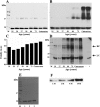
Figure 1
Figure 1
Detection of K2-acetylated γD-crystallin in the human lens. Western blot analysis of γD-crystallin and Nε-acetyllysine-modified proteins in the human lens. Water-soluble human lens proteins were subjected to Western blot analysis using a monoclonal antibody against γD-crystallin (A). The membrane was stripped and reprobed using a monoclonal antibody against Nε-acetyllysine (B). Densitometry of Western blot B is shown in C. Water-soluble human lens proteins were immunoprecipitated using a monoclonal antibody against γD-crystallin and were subjected to Western blot analysis using an antibody against Nε-acetyllysine (D). The age of the donor lenses is shown below the lanes. M denotes the molecular weight markers. Arrows indicate the positions of the light (LC) and heavy chains (HC) of the antibody. The (−) denotes nonacetylated recombinant γD-crystallin; the (+) denotes in vitro acetylated recombinant γD-crystallin. SDS-PAGE of the purified γD-crystallin is shown in panel E. Lanes 1 and 2 are two preparations of γD-crystallin, and lane 3 is in vitro acetylated γD-crystallin. Western blot analysis of acetylated γD-crystallin using an antibody against Nε-acetyllysine; acetylation was carried out using various molar excess concentrations of Ac2O relative to lysine in γD-crystallin (F).
Figure 2
Figure 2
Mass spectrometric detection of acetylation at G1 and K2 in human γD-crystallin. Tandem mass spectra of γD-crystallin from a 73-year-old human lens (A) and in vitro acetylated γD-crystallin (B). The precursor ion of 589.81 (2+) that indicates a mass shift of +84 Da compared with the unmodified peptide is shown. The mass shift of +42 Da was observed at y8, but not y-series ions from y1 to y7, which indicated acetylation of K2. The mass shift of +84 Da was observed on the precursor ion, as well as b-series ions from b2 to b7, which suggested acetylation of K2 and G1.
Figure 3
Figure 3
Acetylated γD-crystallin is more prone to thermal aggregation and less protected by αL-crystallin. (A) Time-course aggregation of nonacetylated and acetylated human γD-crystallin at 80 °C. The samples were prepared in 10 mM phosphate buffer containing 1 mM EDTA (pH 7.0). 1: Nonacetylated; 2: acetylated; 3: nonacetylated with 5 mM DTT; and 4: acetylated with 5 mM DTT. The protein concentration was 0.1 mg/mL. (B) Scattering values of both proteins during thermal aggregation after 1 h. (C) Time-course aggregation of nonacetylated and acetylated γD-crystallin at 80 °C in the presence/absence of αL-crystallin. The samples were prepared in 10 mM phosphate buffer containing 5 mM DTT and 1 mM EDTA (pH 7). The concentration of both γD-crystallin proteins was 0.1 mg/mL. 1: Nonacetylated; 2: acetylated; 3: αL-crystallin alone; 4: nonacetylated + αL-crystallin (1:1) (w/w); 5: acetylated + αL-crystallin (1:1) (w/w); 6: nonacetylated + αL-crystallin (1:0.75) (w/w); and 7: acetylated + αL-crystallin (1:0.75)(w/w). (D) Percent protection of nonacetylated and acetylated γD-crystallin by αL-crystallin during thermal aggregation after 1 h. Bars represent the means ± SD of three independent experiments. *p < 0.05 and ***p < 0.0005.
Figure 4
Figure 4
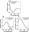
Acetylation perturbed only the tertiary structure of γD-crystallin. (A) Far-UV CD spectra of nonacetylated and acetylated human γD-crystallin. (B) Near-UV CD spectra of nonacetylated and acetylated human γD-crystallin. The concentrations of the protein samples used in far- and near-UV CD were 0.2 and 1.0 mg/mL, respectively. (C) Intrinsic tryptophan fluorescence spectra of nonacetylated and acetylated human γD-crystallin (0.025 mg/mL) were recorded from 310 to 400 nm. The excitation wavelength was 295 nm. Excitation and emission slit widths were 5 nm each. The data were collected at a 0.5 nm wavelength resolution. All assays were performed in 10 mM phosphate buffer containing 1 mM EDTA and 5 mM DTT (pH 7.0) at 25 °C.
Figure 5
Figure 5
Conformational analysis of nonacetylated and acetylated γD-crystallin using molecular dynamics simulations. Molecular dynamics simulation results suggest that acetylation does not alter the secondary structure of γD-crystallin (A). Tryptophan residues in the nonacetylated (yellow), in the K2-acetylated (orange) and in the G1- and K2-acetylated γD-crystallin (blue) models are shown (B). The structures used for superposition are models from the top conformational cluster. All tryptophan residues were found to be buried within the globular core of the proteins. The “circled” portions in the panel B represents modulation in the structure/conformation of human γD-crystallin due to aceylation.
Figure 6
Figure 6
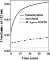
Acetylation alters the cysteine microenvironment in γD-crystallin. DTNB reaction kinetic profiles of nonacetylated and acetylated γD-crystallin at 25 °C. A protein concentration of 0.1 mg/mL in 50 mM phosphate buffer containing 1 mM EDTA (pH 7.4) was used. DTNB was used at a 7-fold molar excess of the protein. Absorbance was measured at 412 nm as a function of time. Mycobacterium leprae HSP18, which lacks cysteine residues, was used as a negative control.
Figure 7
Figure 7
Orientation of thiol residues in nonacetylated and acetylated γD-crystallin. The relative distance between C18 and C78 in the nonacetylated (A), K2-acetylated (B), and G1- and K2-acetylated γD-crystallin (C) calculated from the top conformational cluster models. Heat map analysis for C18 and C78 of human γD-crystallin [nonacetylated (D), K2-acetylated (E), and G1- and K2-acetylated γD-crystallin (F)] was prepared using the root-mean-square deviation of C18 and C78 as collective variables for the y-axis and the distance between C18 and C78 as the collective variable for the x-axis.
Figure 8
Figure 8
Surface electrostatic potential attributable to acetylation in human γD-crystallin. Molecular electrostatic potential surfaces for nonacetylated (A), K2-acetylated (D), and G1- and K2-acetylated (G) human γD-crystallin that were obtained using the adaptive Poisson–Boltzmann solver. Blue and red contours represent electropositive and electronegative isosurfaces at ±0.3 kT/e, respectively. The residues within a 4-Å radius of K2 (green) and G1 (cyan) are represented as sticks with hydrogens, and hydrogen bonds are represented as red dotted lines (B, E, and H). The vacuum-generated electrostatic potentials near the acetylation region are highlighted in all models, and the models show the electropositive (nonacetylated: C) and electronegative potentials (K2 acetylation: F; G1 and K2 acetylation: I). The yellow arrow in panel D, F, G, and I depicts comparative changes due to acetylation of human γD-crystallin.
Figure 9
Figure 9
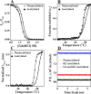
Acetylated γD-crystallin is structurally less stable than the nonacetylated γD-crystallin. Equilibrium GdnHCl unfolding profile for 0.025 mg/mL of nonacetylated and acetylated human γD-crystallin at 37 °C (A). The profile was normalized to a scale of 0–1. Symbols represent the experimental data points, and the solid lines represent the best fit according to the three-state model. The thermal stability of both proteins was evaluated by far-UV CD measurements (B). The temperature was controlled by a water bath, and the data were recorded at a given temperature after a 2 min equilibration. A protein concentration of 0.1 mg/mL in 10 mM phosphate buffer, 5 mM DTT, and 1 mM EDTA (pH 7.0) was used. The raw data were fitted to a two-state model, and the fitting results are represented by solid lines. The thermal stability of both proteins was evaluated by monitoring the intrinsic tryptophan fluorescence (C). The temperature was controlled by a water bath, and the data were recorded at a given temperature after a 2 min equilibration. A protein concentration of 0.025 mg/mL in 10 mM phosphate buffer, 5 mM DTT, and 1 mM EDTA (pH 7.0) was used. The raw data were fitted to a two-state model, and the fitting results are represented by solid lines. Potential energy estimation computed from the molecular dynamics simulation (D). The relative difference in the potential energy profiles indicate the stability of the macro-models in the order nonacetylated > K2-acetylated > G1- and K2-acetylated.
Figure 10
Figure 10
Acetylation alters the unfolding and refolding of γD-crystallin. Productive kinetic unfolding data of nonacetylated and acetylated human γD-crystallin (A). For unfolding, nonacetylated proteins (0.1 mg/mL) were diluted into 5.5 M GdnHCl, 10 mM phosphate buffer, 5 mM DTT, and 1 mM EDTA (pH 7.0) at 25 °C. Changes in the fluorescence intensity at 355 nm were monitored over time using an excitation wavelength of 295 nm. The final protein concentration in the unfolding buffer was 0.01 mg/mL. Unfolding time-course profiles (B) of both proteins were fitted with double and single exponentials, respectively, as indicated by solid lines. Productive kinetic refolding data of nonacetylated and acetylated human γD-crystallin (C). The protein (0.1 mg/mL) was initially unfolded in 5.5 M GdnHCl and diluted into 10 mM phosphate, 5 mM DTT, and 1 mM EDTA (pH 7.0) at 25 °C to yield a final GdnHCl concentration of 1.0 M. The final protein concentration was 0.01 mg/mL. Changes in the fluorescence intensity at 355 nm were monitored over time using an excitation wavelength of 295 nm. Both refolding time-course profiles were fitted with double exponentials, as indicated by solid lines (D).
Figure 11
Figure 11
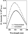
Acetylation exposes additional hydrophobic sites at the surface of γD-crystallin. The concentrations of the protein samples and bis-ANS were 2.5 μM and 10 μM, respectively. All samples were prepared in 10 mM phosphate buffer, 5 mM DTT, and 1 mM EDTA (pH 7.0). The fluorescence spectrum of bis-ANS bound to different samples was recorded from 450 to 600 nm at 25 °C. The excitation wavelength was 390 nm.

References

    1. Bloemendal H. (1982) Lens proteins. CRC Crit. Rev. Biochem. 12, 1–38. - PubMed
    1. Benedek G. B. (1971) Theory of transparency of the eye. Appl. Opt. 10, 459–473. - PubMed
    1. Delaye M.; Tardieu A. (1983) Short-range order of Crystallin proteins accounts for eye lens transparency. Nature 302, 415–417. - PubMed
    1. Bron A. J.; Vrensen G. F.; Koretz J.; Maraini G.; Harding J. J. (2000) The ageing lens. Ophthalmologica 214, 86–104. - PubMed
    1. Horwitz J. (1992) Alpha-Crystallin can function as a molecular chaperone. Proc. Natl. Acad. Sci. U. S. A. 89, 10449–10453. - PMC - PubMed

Publication types