

Application of 3-dimensional printing technology to construct an eye model for fundus viewing study
- PMID: 25393277
- PMCID: PMC4230932
- DOI: 10.1371/journal.pone.0109373
Application of 3-dimensional printing technology to construct an eye model for fundus viewing study
Abstract
Objective: To construct a life-sized eye model using the three-dimensional (3D) printing technology for fundus viewing study of the viewing system.
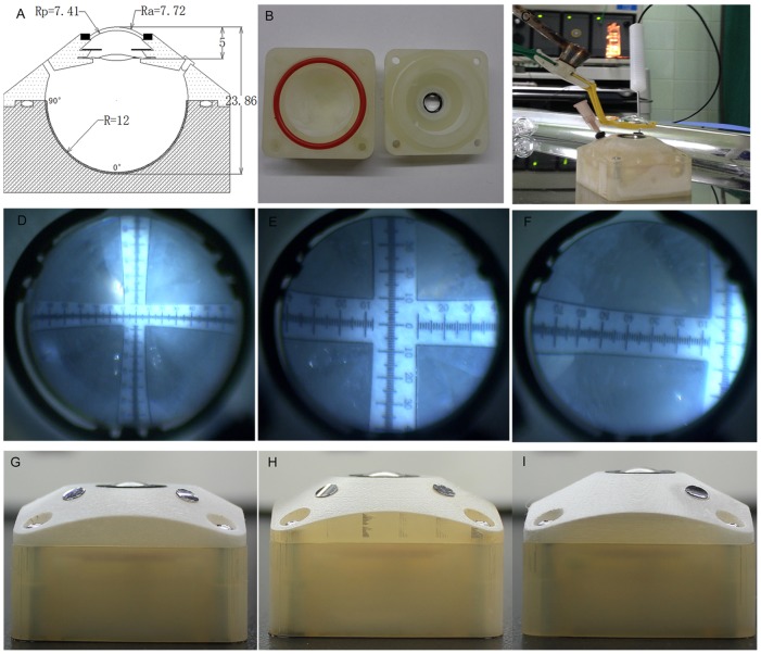
Methods: We devised our schematic model eye based on Navarro's eye and redesigned some parameters because of the change of the corneal material and the implantation of intraocular lenses (IOLs). Optical performance of our schematic model eye was compared with Navarro's schematic eye and other two reported physical model eyes using the ZEMAX optical design software. With computer aided design (CAD) software, we designed the 3D digital model of the main structure of the physical model eye, which was used for three-dimensional (3D) printing. Together with the main printed structure, polymethyl methacrylate(PMMA) aspherical cornea, variable iris, and IOLs were assembled to a physical eye model. Angle scale bars were glued from posterior to periphery of the retina. Then we fabricated other three physical models with different states of ammetropia. Optical parameters of these physical eye models were measured to verify the 3D printing accuracy.
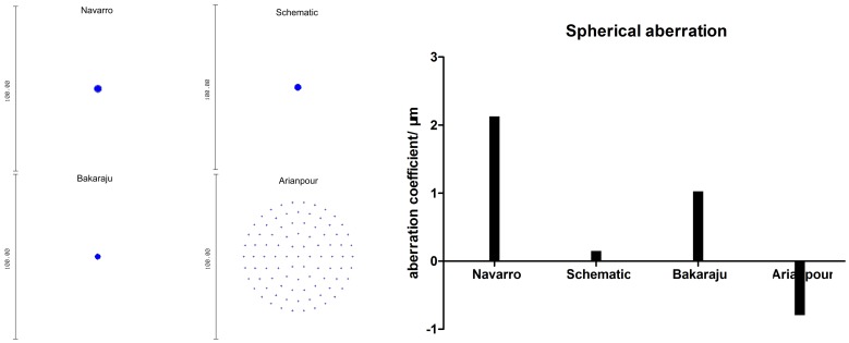
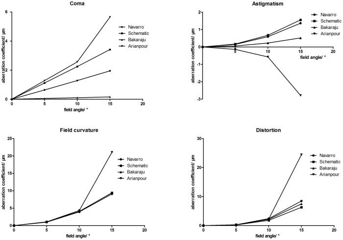
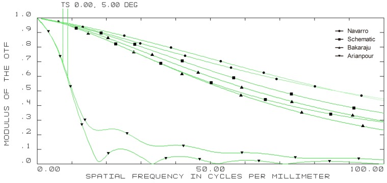
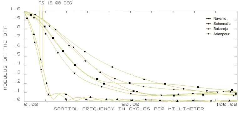
Results: In on-axis calculations, our schematic model eye possessed similar size of spot diagram compared with Navarro's and Bakaraju's model eye, much smaller than Arianpour's model eye. Moreover, the spherical aberration of our schematic eye was much less than other three model eyes. While in off- axis simulation, it possessed a bit higher coma and similar astigmatism, field curvature and distortion. The MTF curves showed that all the model eyes diminished in resolution with increasing field of view, and the diminished tendency of resolution of our physical eye model was similar to the Navarro's eye. The measured parameters of our eye models with different status of ametropia were in line with the theoretical value.
Conclusions: The schematic eye model we designed can well simulate the optical performance of the human eye, and the fabricated physical one can be used as a tool in fundus range viewing research.
Conflict of interest statement
Figures


References
-
- Gullstrand A (1909) The optical system of the eye. In: Helmholtz H, editor. Physioloyische Optik. pp.350–358.
-
- Kooijman AC (1983) Light distribution on the retina of a wide-angle theoretical eye. J Opt Soc Am A 7311: 1544–1550. - PubMed
-
- Navarro R, Santamaría J, Bescós J (1985) Accommodation-dependent model of the human eye with aspherics. J Opt Soc Am A 28: 1273–1280. - PubMed
-
- Escudero-Sanz I, Navarro R (1999) Off-axis aberrations of a wide-angle schematic eye model. J Opt Soc Am A 168: 1881–1891. - PubMed
-
- Gobbi PG, Fasce F, Bozza S, Brancato R (2006) Optomechanical eye model with imaging capabilities for objective evaluation of intraocular lenses. J Cataract Refract Surg A 32: 643–651. - PubMed
Publication types
MeSH terms
Substances
LinkOut - more resources
Full Text Sources
Other Literature Sources
Miscellaneous
