. 2015 Jan 7;54(2):476-480.
doi: 10.1002/anie.201407946. Epub 2014 Nov 13.

Biocompatible infinite-coordination-polymer nanoparticle-nucleic-acid conjugates for antisense gene regulation

Affiliations

Biocompatible infinite-coordination-polymer nanoparticle-nucleic-acid conjugates for antisense gene regulation

Colin M Calabrese et al. Angew Chem Int Ed Engl. .

Abstract

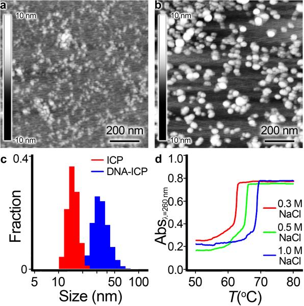
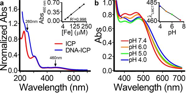
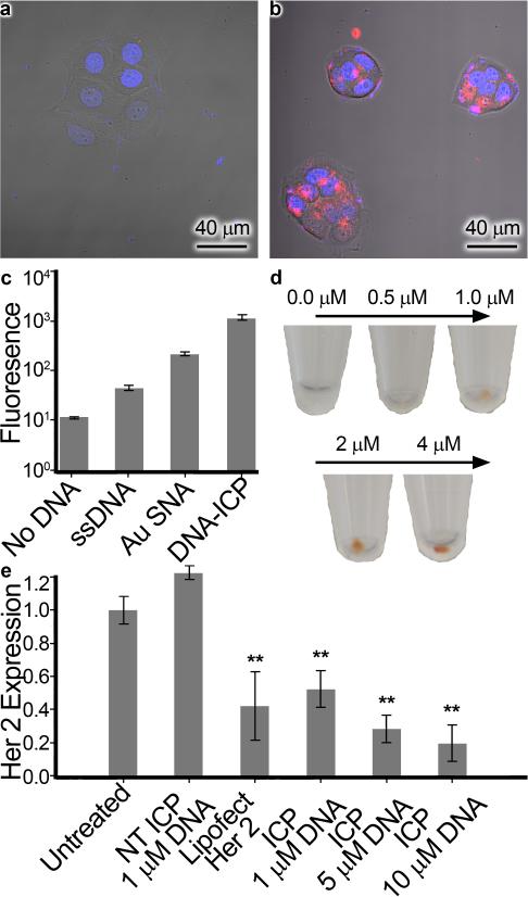
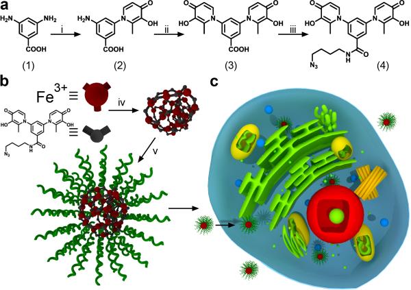
Herein, we report the synthesis of DNA-functionalized infinite-coordination-polymer (ICP) nanoparticles as biocompatible gene-regulation agents. ICP nanoparticles were synthesized from ferric nitrate and a ditopic 3-hydroxy-4-pyridinone (HOPO) ligand bearing a pendant azide. Addition of Fe(III) to a solution of the ligand produced nanoparticles, which were colloidally unstable in the presence of salts. Conjugation of DNA to the Fe(III)-HOPO ICP particles by copper-free click chemistry afforded colloidally stable nucleic-acid nanoconstructs. The DNA-ICP particles, when cross-linked through sequence-specific hybridization, exhibited narrow, highly cooperative melting transitions consistent with dense DNA surface loading. The ability of the DNA-ICP particles to enter cells and alter protein expression was also evaluated. Our results indicate that these novel particles carry nucleic acids into mammalian cells without the need for transfection agents and are capable of efficient gene knockdown.

Keywords: antisense gene regulation; coordination polymers; gene knockdown; iron; nanoparticles.


Figures

Figure 1
Figure 1
Characterization of DNA-ICP particles. AFM image of (a) Bare ICP particles drop-cast and dried on mica. b) DNA-functionalized ICP particles drop-cast and dried on mica. c) DLS histograms comparing size distributions of bare and DNA-functionalized ICPs. d) Cooperative melting of ICP-DNA aggregates.
Figure 2
Figure 2
UV-Vis analysis of DNA-ICP particles. a) Comparison of bare ICP particles with DNA-ICP particles showing the DNA absorbance at 260 nm. Inset: determination of LMCT ε460. b) pH dependence of LMCT absorbance. The red-shift of λmax with decreasing pH is indicative of complex dissociation (see inset).
Figure 3
Figure 3
Cellular uptake and gene knockdown. Confocal microscopy image of HeLa cells treated with (a) Cy5-ssDNA and (b) DNA-ICP particles (100 nM DNA in each case). Hoechst stain denotes the nucleus in blue while the Cy5 dye attached to the DNA is red. c) Fluorescence intensity of Cy5 dye quantified by flow cytometry. d) Naked-eye visualization of DNA-ICPs taken up in pelleted SKOV-3 ovarian cancer cells e) Expression of HER2 protein in SKOV-3 cells treated with non-targeting DNA-ICPs, HER2 targeting ssDNA + Lipofectamine (25 nM DNA basis), and HER2 targeting DNA-ICPS. Starred bars (**) indicate knockdown was significant (p<0.05) as determined by unpaired student's T test.
Scheme 1
Scheme 1
Synthesis and assembly of ICP particles and their cellular uptake. a) Synthetic scheme for bis-3,4-HOPO azide (4). b) Assembly of ICP particles from Fe(NO3)3 and compound 4, followed by conjugation with DNA via a Cu free ‘Click’ reaction. c) Scheme depicting the cellular uptake of ICP-DNA conjugates. Reaction conditions: i) maltol, n-propanol, reflux, 16 h. ii) maltol, ethoxyethanol, 64 h, reflux. iii) 4-azido-butan-1-amine, HATU, diisopropylethylamine, DMSO, RT, 4 h. iv) Fe(NO3)3·9H2O, NaOH (aq.), RT, 10 min. v) Dibenzocyclooctyne-DNA, 0.5M NaCl, RT, 16 h

References

    1. Macfarlane RJ, Lee B, Jones MR, Harris N, Schatz GC, Mirkin CA. Science. 2011;334:204–208. - PubMed
    2. Nykypanchuk D, Maye MM, van der Lelie D, Gang O. Nature. 2008;451:549–552. - PubMed
    1. Zheng D, Seferos DS, Giljohann DA, Patel PC, Mirkin CA. Nano Lett. 2009;9:3258–3261. - PMC - PubMed
    2. Seferos DS, Giljohann DA, Hill HD, Prigodich AE, Mirkin CA. J. Am. Chem. Soc. 2007;129:5477. - PMC - PubMed
    3. Prigodich AE, Randeria PS, Briley WE, Kim NJ, Daniel WL, Giljohann DA, Mirkin CA. Anal. Chem. 2012;84:2062–2066. - PMC - PubMed
    1. Rosi NL, Giljohann DA, Thaxton CS, Lytton-Jean AKR, Han MS, Mirkin CA. Science. 2006;312:1027–1030. - PubMed
    2. Giljohann DA, Seferos DS, Prigodich AE, Patel PC, Mirkin CA. J. Am. Chem. Soc. 2009;131:2072. - PMC - PubMed
    1. Cutler JI, Zhang K, Zheng D, Auyeung E, Prigodich AE, Mirkin CA. J. Am. Chem. Soc. 2011;133:9254–9257. - PMC - PubMed
    2. Young KL, Scott AW, Hao L, Mirkin SE, Liu G, Mirkin CA. Nano Lett. 2012;12:3867–3871. - PMC - PubMed
    1. Mirkin CA, Letsinger RL, Mucic RC, Storhoff JJ. Nature. 1996;382:607–609. - PubMed

Publication types