

Contribution of cone beam computed tomography to the detection of apical root resorption after orthodontic treatment in root-filled and vital teeth
- PMID: 25393801
- PMCID: PMC8610389
- DOI: 10.2319/042814-308.1
Contribution of cone beam computed tomography to the detection of apical root resorption after orthodontic treatment in root-filled and vital teeth
Abstract
Objective: To investigate whether root-filled teeth are similar to vital pulp teeth in terms of apical root resorption (ARR) after orthodontic treatment.
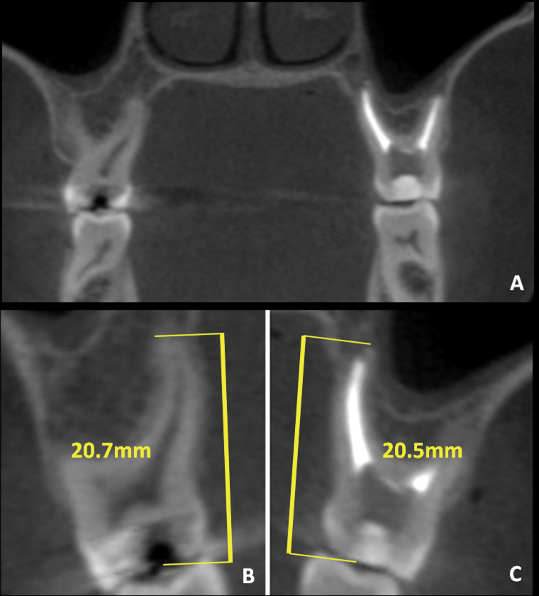
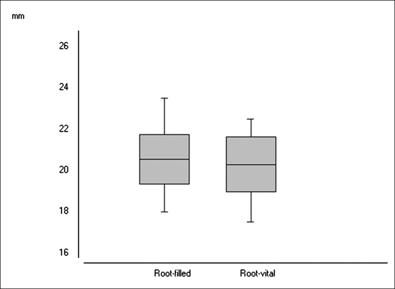
Materials and methods: An original sample of cone beam computed tomography (CBCT) images of 1256 roots from 30 orthodontic patients were analyzed. The inclusion criteria demanded root-filled teeth and their contralateral vital teeth, while teeth with history of trauma had to be excluded to comply with exclusion criteria. CBCT images of root-filled teeth were compared before and after orthodontic treatment in a split-mouth design study. Tooth measurements were made with multiplanar reconstruction using axial-guided navigation. The statistical difference between the treatment effects was compared using the paired t-test.
Results: Twenty posterior root-filled teeth and their contralaterals with vital pulp were selected before orthodontic treatment from six adolescents (two boys and four girls; mean [SD] age 12.8 [1.8] years). No differences were detected between filled and vital root lengths before treatment (P = .4364). The mean differences in root length between preorthodontic and postorthodontic treatment in filled- and vital roots were -0.30 mm and -0.16 mm, respectively, without any statistical difference (P = .4197) between them.
Conclusion: There appears to be no increase in ARR after orthodontic treatment in root-filled teeth with no earlier ARR.
Keywords: Cone beam computed tomography; Endodontics; Orthodontics; Root resorption.
Figures
References
-
- Levander E, Malmgren O. Evaluation of the risk of root resorption during orthodontic treatment: a study of upper incisors. Eur J Orthod. 1988;10:30–38. - PubMed
-
- Brudvik P, Rygh P. Multi-nucleated cells remove the main hyalinized tissue and start resorption of adjacent root surfaces. Eur J Orthod. 1994;16:265–273. - PubMed
-
- Sameshima GT, Sinclair PM. Predicting and preventing root resorption: Part II. Treatment factors. Am J Orthod Dentofacial Orthop. 2001;119:511–515. - PubMed
-
- Malmgren O, Goldson L, Hill C, Orwin A, Petrini L, Lundberg M. Root resorption after orthodontic treatment of traumatized teeth. Am J Orthod Dentofacial Orthop. 1982;82:487–491. - PubMed
MeSH terms
LinkOut - more resources
Full Text Sources
Other Literature Sources
