. 2015 Feb;37(2):182-94.
doi: 10.1002/bies.201400145. Epub 2014 Nov 13.

Multiple dimensions of epigenetic gene regulation in the malaria parasite Plasmodium falciparum: gene regulation via histone modifications, nucleosome positioning and nuclear architecture in P. falciparum

Affiliations

Multiple dimensions of epigenetic gene regulation in the malaria parasite Plasmodium falciparum: gene regulation via histone modifications, nucleosome positioning and nuclear architecture in P. falciparum

Ferhat Ay et al. Bioessays. 2015 Feb.

Abstract

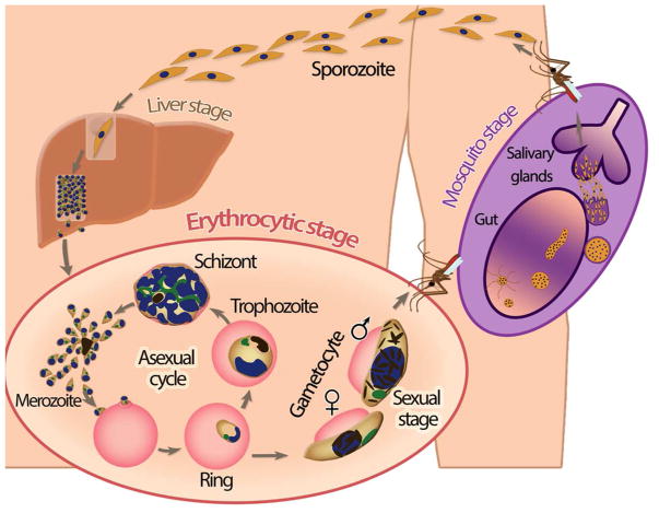
Plasmodium falciparum is the most deadly human malarial parasite, responsible for an estimated 207 million cases of disease and 627,000 deaths in 2012. Recent studies reveal that the parasite actively regulates a large fraction of its genes throughout its replicative cycle inside human red blood cells and that epigenetics plays an important role in this precise gene regulation. Here, we discuss recent advances in our understanding of three aspects of epigenetic regulation in P. falciparum: changes in histone modifications, nucleosome occupancy and the three-dimensional genome structure. We compare these three aspects of the P. falciparum epigenome to those of other eukaryotes, and show that large-scale compartmentalization is particularly important in determining histone decomposition and gene regulation in P. falciparum. We conclude by presenting a gene regulation model for P. falciparum that combines the described epigenetic factors, and by discussing the implications of this model for the future of malaria research.

Keywords: epigenetics; gene regulation; histone modifications; malaria; nucleosome occupancy; three-dimensional genome organization; virulence genes.


Conflict of interest statement

The authors have declared no conflict of interest.

Figures

Figure 1
Figure 1
Overview of the P. falciparum life cycle.
Figure 2
Figure 2. Large-scale depletion of the transcriptionally permissive histone variant H2A.Z and activating histone marks in the telomeric cluster visualized on the 3D P. falciparum genome
ChIP-seq data from Bartfai et al. [45] for 4 histone variants or marks were downloaded from GEO (accession number: GSE23787) and mapped to the P. falciparum genome (PlasmoDB v9.0) using the short read alignment mode of BWA (v0.5.9) [117] with default parameter settings. Reads were post-processed, and only the reads that map uniquely with a quality score above 30 and with at most two mismatches were retained for further analysis. Retained reads were subjected to PCR duplicate elimination and then were aggregated for each non-overlapping 5 kb bin across the P. falciparum genome. The number of reads for each 5 kb bin was normalized using the overall sequencing depth of the corresponding ChIP-seq library. Plotted are the log2 ratios of sequence-depth normalized number of reads from the ChIP-seq library versus the corresponding input library (red: depletion, blue: enrichment) for A: H2A at 40 hours post invasion (hpi), B: H2A.Z at 10 hpi, C: H2A.Z at 30 hpi, D: H2A.Z at 40 hpi, E: H3K9ac at 40 hpi, and F: H3K4me3 at 40 hpi. 3D models for the ring, trophozoite and schizont stages were generated in Ay et al. [30] and were colored with ChIP-seq enrichment/depletion from 10, 20 and 40 hpi, respectively. Light blue and white spheres indicate centromeres and telomeres, respectively. The black dashed circle denotes the telomeric cluster for each stage. See http://noble.gs.washington.edu/proj/plasmo-epigenetics for the rotating 3D figure of each available ChIP-seq library.
Figure 3
Figure 3. Enrichment of repressive histone marks in the telomeric cluster visualized on the 3D P. falciparum genome at the ring stage
ChIP-seq data from Jiang et al. [46] for 5 histone marks were downloaded from SRA (accession number: SRP022761) and processed as described in the caption of Figure 2. Due to lack of input libraries from this publication, the input libraries from Bartfai et al. at different time points were pooled into one aggregated input library which was then used for normalization of each Jiang et al. ChIP-seq library. Similar to Figure 2, log2 ratios of ChIP-seq versus input were plotted for A: H3K9me3, B: H3K36me3, C: H4K20me3, and D: H3K4me3 at 18 hpi. The 3D model for the ring stage from Ay et al. [30] was used to visualize enrichment/depletion of each histone mark. See http://noble.gs.washington.edu/proj/plasmo-epigenetics for the rotating 3D figure of each available ChIP-seq library.
Figure 4
Figure 4. Model for P. falciparum epigenetic gene regulation
A: Nuclear organization and gene regulation in P. falciparum. Centromeric (dark blue) and telomeric (red) clusters are localized at the nuclear periphery. Subtelomeric virulence genes (blue) are anchored to the nuclear perimeter and cluster with internally located var genes in repressive center(s), characterized by repressive histone marks H3K9me3 and H3K36me3. The single active var gene (green) is located in a perinuclear compartment away from the repressive center(s). In addition, active rDNA genes (orange) also cluster at the nuclear periphery. The remaining genome (purple) is largely present in an open, euchromatic state with a number of notable features. (i) Nucleosome levels are high in genic and lower in intergenic regions, while gene expression correlates with nucleosome density at the transcription start site. (ii) Intergenic regions are bound by nucleosomes containing histone variants H2A.Z and H2B.Z. (iii) Intergenic regions contain H3K4me3, the level of which does not influence transcriptional activity. (iv) H3K9ac is mainly found in intergenic regions and extends into 5′ ends of coding regions, with highly expressed genes showing higher levels of H3K9ac. (v) Active genes are marked with H3K36me3 towards their 3′ end. B: Remodeling of the nuclear organization during the asexual cycle. Extensive remodeling of the nucleus takes place as the parasite progresses through the ring, trophozoite and schizont stages. In the transition from the relatively inert ring stage to the transcriptionally active trophozoite stage, the size of the nucleus and the number of nuclear pores increase, accompanied by a decrease in genome-wide nucleosome levels, resulting in an open chromatin structure that allows high transcription rates. In the schizont stage, the nucleus divides and recompacts, histones are re-assembled and transcription is shutdown, to facilitate egress of the parasites’ daughter cells and invasion of new red blood cells.

References

    1. Greenwood BM, Fidock DA, Kyle DE, Kappe SH, et al. Malaria: progress, perils, and prospects for eradication. The Journal of clinical investigation. 2008;118:1266–76. - PMC - PubMed
    1. Baker DA. Malaria gametocytogenesis. Mol Biochem Parasitol. 2010;172:57–65. - PMC - PubMed
    1. Baruch DI, Pasloske BL, Singh HB, Bi X, et al. Cloning the P. falciparum gene encoding PfEMP1, a malarial variant antigen and adherence receptor on the surface of parasitized human erythrocytes. Cell. 1995;82:77–87. - PubMed
    1. Smith JD, Chitnis CE, Craig AG, Roberts DJ, et al. Switches in expression of Plasmodium falciparum var genes correlate with changes in antigenic and cytoadherent phenotypes of infected erythrocytes. Cell. 1995;82:101–10. - PMC - PubMed
    1. Su XZ, Heatwole VM, Wertheimer SP, Guinet F, et al. The large diverse gene family var encodes proteins involved in cytoadherence and antigenic variation of Plasmodium falciparum-infected erythrocytes. Cell. 1995;82:89–100. - PubMed

Publication types

MeSH terms