. 2014 Dec 11;371(24):2277-2287.
doi: 10.1056/NEJMoa1409354. Epub 2014 Nov 13.

Thrombospondin type-1 domain-containing 7A in idiopathic membranous nephropathy

Affiliations

Thrombospondin type-1 domain-containing 7A in idiopathic membranous nephropathy

Nicola M Tomas et al. N Engl J Med. .

Abstract

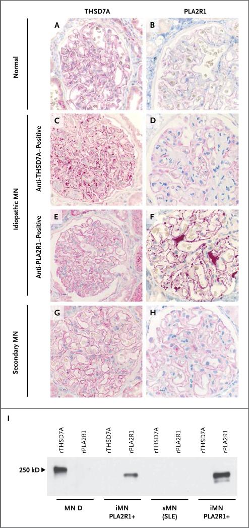
Background: Idiopathic membranous nephropathy is an autoimmune disease. In approximately 70% of patients, it is associated with autoantibodies against the phospholipase A2 receptor 1 (PLA2R1). Antigenic targets in the remaining patients are unknown.

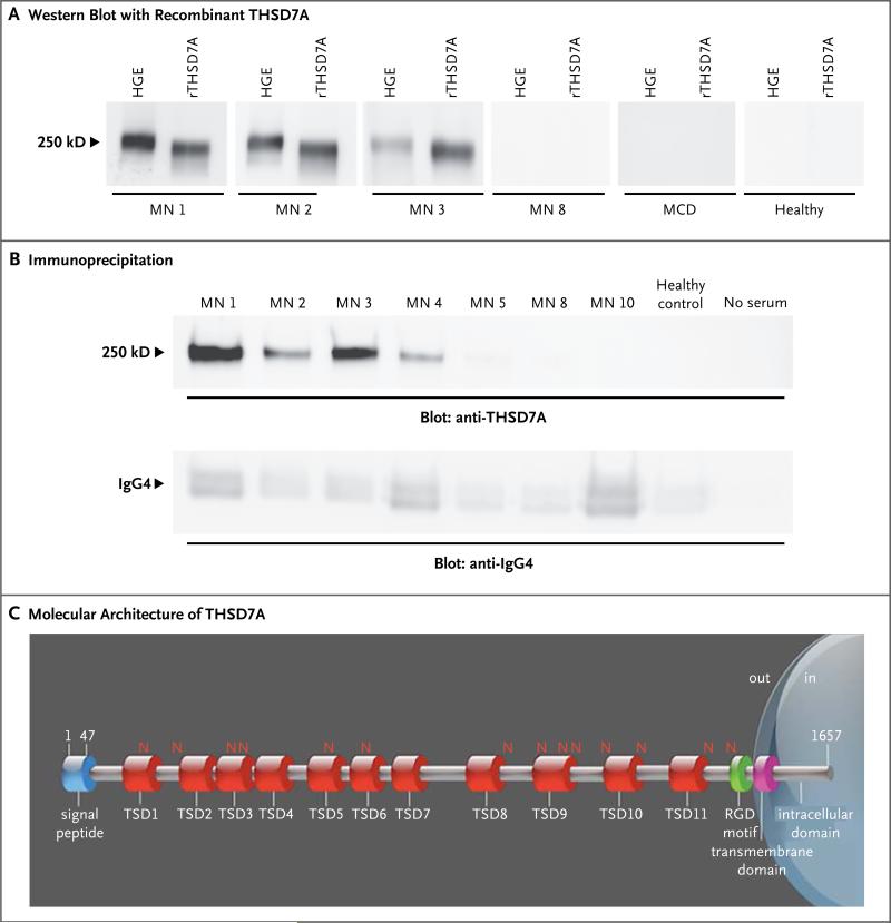
Methods: Using Western blotting, we screened serum samples from patients with idiopathic membranous nephropathy, patients with other glomerular diseases, and healthy controls for antibodies against human native glomerular proteins. We partially purified a putative new antigen, identified this protein by means of mass spectrometry of digested peptides, and validated the results by analysis of recombinant protein expression, immunoprecipitation, and immunohistochemical analysis.

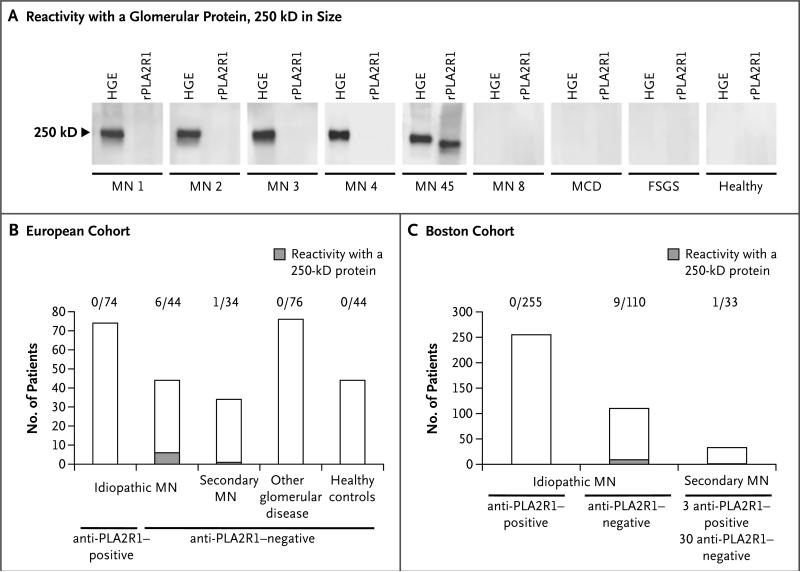
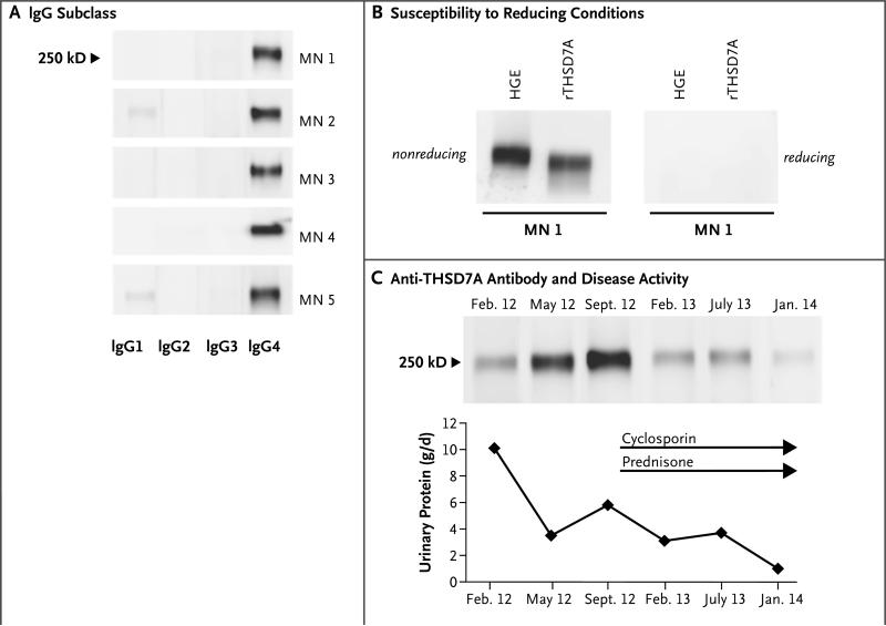
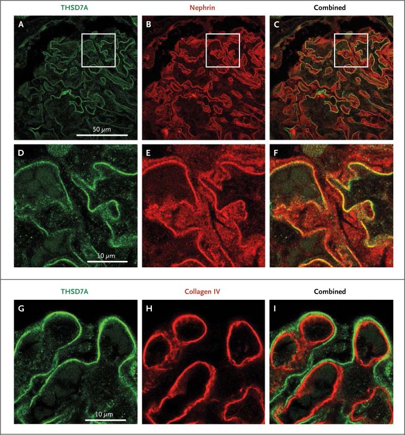
Results: Serum samples from 6 of 44 patients in a European cohort and 9 of 110 patients in a Boston cohort with anti-PLA2R1-negative idiopathic membranous nephropathy recognized a glomerular protein that was 250 kD in size. None of the serum samples from the 74 patients with idiopathic membranous nephropathy who were seropositive for anti-PLA2R1 antibodies, from the 76 patients with other glomerular diseases, and from the 44 healthy controls reacted against this antigen. Although this newly identified antigen is clearly different from PLA2R1, it shares some biochemical features, such as N-glycosylation, membranous location, and reactivity with serum only under nonreducing conditions. Mass spectrometry identified this antigen as thrombospondin type-1 domain-containing 7A (THSD7A). All reactive serum samples recognized recombinant THSD7A and immunoprecipitated THSD7A from glomerular lysates. Moreover, immunohistochemical analyses of biopsy samples from patients revealed localization of THSD7A to podocytes, and IgG eluted from one of these samples was specific for THSD7A.

Conclusions: In our cohort, 15 of 154 patients with idiopathic membranous nephropathy had circulating autoantibodies to THSD7A but not to PLA2R1, a finding that suggests a distinct subgroup of patients with this condition. (Funded by the French National Center for Scientific Research and others.).


Figures

Figure 1
Figure 1. Detection of Autoantibodies against an Unknown Antigen in Patients with Membranous Nephropathy
Panel A shows reactivity of serum samples with human glomerular extracts (HGE). Four of the initially tested samples from patients with membranous nephropathy (MN) reacted with a glomerular protein approximately 250 kD in size (MN 1 through MN 4) but not with the recombinant phospholipase A2 receptor (rPLA2R1). Panels B and C show overall reactivity in the European and Boston cohorts, respectively. FSGS denotes focal segmental glomerulo-sclerosis, and MCD minimal-change disease. The number above each column indicates the fraction of samples positive for a 250-kD protein.
Figure 2
Figure 2. Identification of the Target Antigen
Panel A shows reactivity of serum samples with human glomerular extracts (HGE) and recombinant thrombospondin type-1 domain-containing 7A (rTHSD7A). Only samples from patients with membranous nephropathy (MN) that previously recognized the 250-kD protein present in HGE also recognized rTHSD7A (MN 1, MN 2, and MN 3). Panel B shows results of immunoprecipitation experiments. All serum samples from patients with membranous nephropathy that previously recognized rTHSD7A precipitated the antigen from HGE. No immunoprecipitation occurred with serum from healthy controls or with no serum (i.e., with water substituted for serum in the experiment). IgG4 was efficiently immunoprecipitated in all conditions (lower panel). Panel C shows the molecular architecture of THSD7A, with a large extracellular region comprising 11 thrombospondin type-1 domains (TSDs), 14 glycosylation sites (N), and 1 predicted arginine–glycine–aspartic acid (RGD) motif. The scheme was built according to the UniProt accession number Q9UPZ6 for THSD7A and ScanProsite tool from ExPASy.
Figure 3
Figure 3. Characterization of Anti-THSD7A Autoantibodies
Panel A shows that anti-THSD7A autoantibodies are predominantly of the IgG4 subclass. Other IgG subtypes were also present in most patients. Panel B shows that serum reactivity against THSD7A is lost in the presence of a reducing agent, a finding that suggests that the autoantibody recognizes a conformation-dependent epitope. Panel C shows results of Western blotting of equal volumes of glomerular proteins with serial serum samples from one patient with idiopathic membranous nephropathy. After an initial lessening of proteinuria, severe edema and severe proteinuria developed in the patient in September 2012. Immunosuppressive treatment with cyclosporine and prednisone induced a partial remission in January 2014. This resolution of disease activity is reflected by a decrease of the Western blot signal after initiation of immunosuppressive treatment.
Figure 4
Figure 4. Expression of THSD7A in Normal Human Glomeruli
Immunofluorescence experiments were performed on renal-biopsy samples from healthy controls who underwent nephrectomy. Panel A shows staining for THSD7A (green) which is found along the glomerular capillary wall in a linear aspect. Panel B shows staining for nephrin (red), a podocyte membrane protein. Panel C shows strong colocalization of THSD7A and nephrin, suggesting that THSD7A is expressed on glomerular podocytes. Panels D, E, and F are enlarged images of the boxed areas in Panels A, B, and C, respectively. Panels G, H, and I show staining for THSD7A, collagen type IV, and both, respectively. THSD7A is localized in an abluminal position relative to collagen type IV, which suggests that it is expressed on glomerular podocytes rather than on the glomerular basement membrane itself.
Figure 5
Figure 5. Expression Level of THSD7A and PLA2R1 in Patients with Idiopathic and Secondary Membranous Nephropathy and in a Healthy Control
Immunohistochemical staining for THSD7A and PLA2R1 was performed in renal-biopsy specimens from healthy controls and from patients with membranous nephropathy. Panels A and B show normal staining for THSD7A and PLA2R1, respectively, in a control biopsy specimen. Panel C shows markedly enhanced staining for THSD7A in a patient with serum anti-THSD7A antibodies, whereas PLA2R1 staining, as shown in Panel D, is normal. Conversely, the biopsy specimen from a patient with serum anti-PLA2R1 antibodies shows that staining for THSD7A was not increased, as shown in Panel E, but staining for PLA2R1 was increased, as shown in Panel F. The biopsy specimen from a patient with secondary membranous nephropathy (sMN) due to systemic lupus erythematosus (SLE) and no detectable serum antibodies against THSD7A or PLA2R1 has normal staining for both antigens, as seen in Panels G and H. Panel I shows the reactivity of IgG eluted from patient biopsy samples with recombinant THSD7A and PLA2R1. Only the IgG eluted from the biopsy sample from a patient with idiopathic membranous nephropathy (iMN) and detectable serum anti-THSD7A antibodies recognized THSD7A (MN D; see also Table S3 in the Supplementary Appendix). The eluates from two biopsy samples from patients with idiopathic membranous nephropathy and serum anti-PLA2R1 autoantibodies recognized PLA2R1 but not THSD7A. IgG eluted from one patient with lupus membranous nephropathy and no detectable serum antibodies against THSD7A or PLA2R1 recognized neither antigen.

Comment in

References

    1. Deegens JK, Wetzels JF. Membranous nephropathy in the older adult: epidemiology, diagnosis and management. Drugs Aging. 2007;24:717–32. - PubMed
    1. Beck LH, Jr, Bonegio RG, Lambeau G, et al. M-type phospholipase A2 receptor as target antigen in idiopathic membranous nephropathy. N Engl J Med. 2009;361:11–21. - PMC - PubMed
    1. Hoxha E, Harendza S, Zahner G, et al. An immunofluorescence test for phospholipase-A₂-receptor antibodies and its clinical usefulness in patients with mem branous glomerulonephritis. Nephrol Dial Transplant. 2011;26:2526–32. - PubMed
    1. Hofstra JM, Beck LH, Jr, Beck DM, Wetzels JF, Salant DJ. Anti-phospholipase A2 receptor antibodies correlate with clinical status in idiopathic membranous nephropathy. Clin J Am Soc Nephrol. 2011;6:1286–91. - PMC - PubMed
    1. Hofstra JM, Debiec H, Short CD, et al. Antiphospholipase A2 receptor antibody titer and subclass in idiopathic membranous nephropathy. J Am Soc Nephrol. 2012;23:1735–43. - PMC - PubMed

Publication types

MeSH terms