. 2014 Nov 14:5:5384.
doi: 10.1038/ncomms6384.

CSN6 drives carcinogenesis by positively regulating Myc stability

Affiliations

CSN6 drives carcinogenesis by positively regulating Myc stability

Jian Chen et al. Nat Commun. .

Abstract

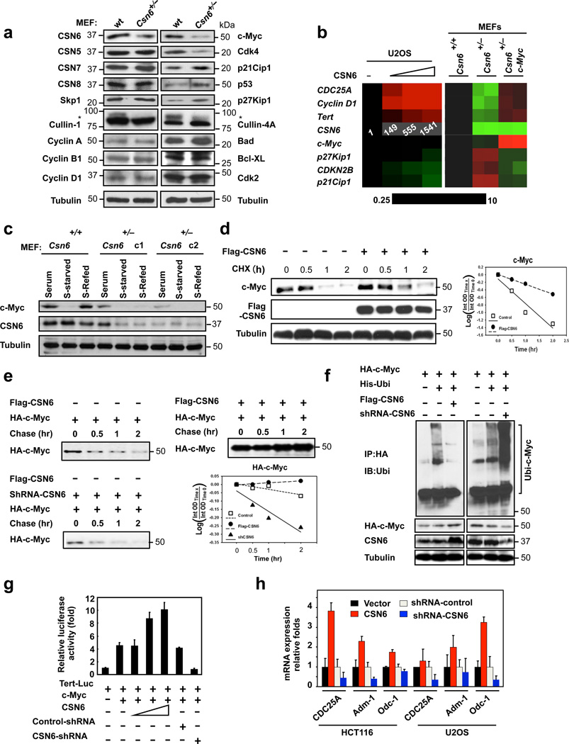
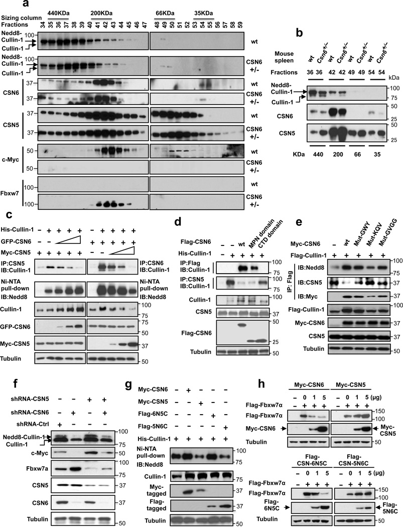
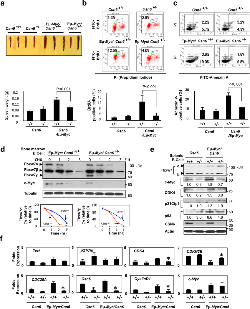
Cullin-RING ubiquitin ligases (CRLs) are critical in ubiquitinating Myc, while COP9 signalosome (CSN) controls neddylation of Cullin in CRL. The mechanistic link between Cullin neddylation and Myc ubiquitination/degradation is unclear. Here we show that Myc is a target of the CSN subunit 6 (CSN6)-Cullin signalling axis and that CSN6 is a positive regulator of Myc. CSN6 enhanced neddylation of Cullin-1 and facilitated autoubiquitination/degradation of Fbxw7, a component of CRL involved in Myc ubiquitination, thereby stabilizing Myc. Csn6 haplo-insufficiency decreased Cullin-1 neddylation but increased Fbxw7 stability to compromise Myc stability and activity in an Eμ-Myc mouse model, resulting in decelerated lymphomagenesis. We found that CSN6 overexpression, which leads to aberrant expression of Myc target genes, is frequent in human cancers. Together, these results define a mechanism for the regulation of Myc stability through the CSN-Cullin-Fbxw7 axis and provide insights into the correlation of CSN6 overexpression with Myc stabilization/activation during tumorigenesis.


Figures

Figure 1
Figure 1. Loss of CSN6 leads to destabilization of Myc and decreases Myc transcriptional activity
(a) Immunoblot analysis of Csn6+/− MEFs. Primary MEF cells were prepared from 13.5-day embryos derived from wild-type (wt) Csn6+/+ and Csn6+/− mice. After 4 days of culture, cell lysates were analyzed by indicated antibodies. (b) Quantitative RT-PCR analysis of mRNA levels for indicated c-Myc target genes in CSN6-transfected U2OS cells or Csn6+/− MEFs. The quantitated mRNA expression level was normalized to GAPDH mRNA. Two MEFs were examined for each genotype. Expression level of transfected CSN6 is indicated as numbers on the heat map, and the heat map depicts the natural logarithm of fold-change in mRNA expression. (c) CSN6 knockdown abrogated serum-induced elevation of Myc. Csn6+/+ MEFs and Csn6+/− clone 1 (C1) and clone 2 (C2) MEFs were serum-starved for 24 hours and then were refed with serum for 24 hours. Lysates were analyzed by indicated antibodies. (d) CSN6 decreased Myc turnover. 293T cells were transfected with Flag-CSN6 and were then treated with cycloheximide (CHX; 100 µg/ml) for the indicated times. The immunoblot of Myc signal at each time point was measured using a densitometer and the integrated optical density of Myc was measured. The turnover of Myc is indicated graphically. (e) CSN6 expression affected Myc turnover. 35S–pulse-labeled HA-Myc protein was immunoprecipitated from indicated transfected 293T cell lysates. The mixture was separated by sodium dodecyl sulfate polyacrylamide electrophoresis (SDS-PAGE) and the gel exposed to x-ray film. The density of HA-Myc was measured and the integrated optical density (OD) was measured. The turnover of HA-Myc is indicated graphically. (f) CSN6 decreased ubiquitination levels of Myc. 293T cells were transfected with indicated plasmids. Cells were treated with 50 µg/ml MG132 for 6 hours before harvest. The ubiquitinated Myc proteins were pulled down with anti-HA antibody and immunoblotted with anti-ubiquitin (Ubi) antibody. (g) CSN6 activated Myc transcriptional activity. The Tert-luc reporter containing a Myc response element (Ebox) was transfected with the Myc–expressing vectors and increasing amounts of the CSN6 expression vector or CSN6 shRNA plasmid into 293T cells. Relative luciferase activities are shown with error bars representing standard deviations. Each result shown is representative of 3 independent experiments. (h) CSN6 elevated expression of Myc target genes. Quantitative real-time RT-PCR analysis of Myc target gene expression, including CDC25A, Adm-1, and ODC-1, in indicated cells. The error bars represent 95% confidence intervals. Each result shown is representative of 3 independent experiments.
Figure 2
Figure 2. CSN6 regulates SCF (Skp1/Cullin-1/Fbxw7) complex to stabilize Myc
(a) CSN6 associated with Myc, Fbxw7 and Cullin-1. The U2OS cell lysates were immunoprecipitated with anti-CSN6 and immunoblotted with indicated antibodies. (b) CSN6 knockdown led to decreased Cullin-1 neddylation. The U2OS cell lysates were immunoprecipitated with anti-Cullin-1, followed by immunoblotting with indicated antibodies. (c) CSN6 expression was inversely related to Fbxw7 expression. Immunoblot analysis of Myc and Fbxw7α in CSN6-overexpressing or CSN6-knockdown cells. Cell lysates were analyzed by indicated antibodies. (d) Knockdown of CSN6 increased the expression of Fbxw7 and reduced Myc. Indicated cells were infected with lentivirus to express one of two independent CSN6 shRNAs (#1, #2) or luciferase control shRNA. Cell lysates were immunoblotted with indicated antibodies. (e) CSN6 reduced steady-state expression of Fbxw7 to upregulate Myc. Indicated cells were transfected with increasing amounts of GFP-CSN6. Cell lysates were immunoblotted with indicated antibodies. Fbxw7α and Fbxw7β isoforms were identified. (h) Knockdown of CSN6 enhanced Myc ubiquitination. Indicated cells were infected with lentivirus to express one of two independent CSN6 shRNAs (#1, #2) or luciferase control shRNA. The cells were then transfected with His-ubiquitin and treated with MG132 for 6 hours before harvest. The ubiquitinated Myc proteins were pulled down using Ni-NTA-agarose beads and detected with anti-Myc antibody.
Figure 3
Figure 3. CSN6 enhances Fbxw7’s autoubiquitination and degradation
(a) CSN6 increased Fbxw7α turnover. 293T cells were infected with lentivirus to express CSN6 shRNA or luciferase control shRNA. The cells were then transfected with Flag-Fbxw7α and were then treated with cycloheximide (CHX; 100 µg/ml) for the indicated times. The density of Fbxw7α was measured and the integrated optical density (OD) was measured. The turnover of Fbxw7α is indicated graphically. (b) CSN6-mediated Fbxw7α downregulation was proteasome-dependent. 293T cells were cotransfected with indicated plasmids and were treated with or without 50 µg/ml MG132 for 6 hours. The cell lysates were then immunoblotted with the indicated antibodies. (c) CSN6 enhanced Fbxw7α ubiquitination through lysine 48 linkage. 293T cells were cotransfected with Flag-Fbxw7α with or without GFP-CSN6 plus His-ubiquitin wild type (wt), K63R mutant, or K48R mutant. Cells were treated with 50 µg/ml MG132 for 6 hours before harvest. The ubiquitinated Fbxw7α proteins were pulled down using Ni-NTA-agarose beads and detected with anti-Flag antibody. (d) An F-box domain deletion mutant of Fbxw7α was resistant to CSN6-mediated ubiquitination. 293T cells were cotransfected with GFP-CSN6, His-ubiquitin plus Flag-Fbxw7α wild-type or F-box domain deletion mutant (ΔF). Cells were treated with 50 µg/ml MG132 for 6 hours before harvest. The ubiquitinated Fbxw7α proteins were pulled down using Ni-NTA-agarose beads and detected with anti-Flag antibody. (e) Cullin-1 knockdown diminished CSN6-induced ubiquitination of Fbxw7α. 293T cells were transfected with siRNA Cullin-1 or siRNA control plus indicated plasmids. Cells were treated with 50 µg/ml MG132 for 6 hours before harvest. The ubiquitinated Fbxw7α proteins were pulled down using Ni-NTA-agarose beads and detected with anti-Flag antibody. (f) Cullin-1 neddylation was involved in CSN6-mediated downregulation of Fbxw7α. 293T cells were transfected with indicated plasmids plus increasing amounts of HA-dnUbc12. The levels of Nedd8-Cullin-1 and Nedd8-dnUbc12 were detected with anti-Nedd8 antibody.
Figure 4
Figure 4. CSN6 enhances neddylation of Cullin-1 via its MPN domain
(a) Neddylation status of Cullin in gel filtration chromatography fractions from spleen extracts from Csn6+/+ (wt) and Csn6+/− mice. Extracts of spleen from CSN6+/+ or Csn6+/− mice (4 weeks old) were ground and subjected to lysis. Lysates were fractionated by gel filtration chromatography. Fractions were resolved by SDS-PAGE, followed by immunoblotting with indicated antibodies. Molecular size of eluted fraction is indicated above. (b) Non-neddylated Cullin was increased in Csn6+/− mice. Extracts of spleen from CSN6+/+ (wt) or Csn6+/− mice were fractionated by gel filtration chromatography. Representative fractions 36–54 from (a) were resolved by SDS-PAGE, followed by immunoblotting with anti-Cullin-1, anti-CSN6, and anti-CSN5 antibodies. (c) CSN6 competed with CSN5 for binding to Cullin-1 and affected Cullin-1 neddylation. 293T cells were transfected with His-Cullin-1 plus increasing amounts of GFP-CSN6 or Myc-CSN5. The neddylated Cullin-1 proteins were pulled down using Ni-NTA-agarose beads and detected with anti-Nedd8 antibody. The lysates were also immunoprecipitated with CSN6 antibody or CSN5 antibody and were subjected to immunoblotting with anti-Cullin-1. (d) MPN domain of CSN6 had an important role in competing with CSN5 for Cullin-1 binding and affected Cullin-1 neddylation. 293T cells were cotransfected with indicated plasmids. The lysates were immunoprecipitated with anti-Flag or anti-CSN5 antibody and immunoblotted with the indicated antibodies. (e) Mutation of conserved residues in the MPN domain of CSN6 compromised CSN6’s capacity for competing with CSN5. 293T cells were cotransfected with indicated CSN6 MPN mutant plasmids. The lysates were immunoprecipitated with anti-Flag antibody and immunoblotted with indicated antibodies. (f) CSN6 antagonized CSN5 in regulating Cullin-1 neddylation. U2OS cells were infected with indicated lentivirus carrying CSN6 shRNA, CSN5 shRNA, or luciferase control shRNA. Cell lysates were immunoblotted with the indicated antibodies. (g) The chimeric protein 6N5C expressing the CSN6 MPN domain functioned like wild-type CSN6 in increasing Cullin-1 neddylation. 293T cells were cotransfected with indicated wild-type– and chimeric protein–expressing plasmids. The lysates were pulled down with Ni-NTA-agarose beads and immunoblotted with anti-Nedd8. (h) Both chimeric protein 6N5C and wild-type CSN6 decreased the stability of Fbxw7α in a dose-dependent manner. 293T cells were cotransfected with indicated plasmids. The lysates were immunoblotted with indicated antibodies.
Figure 5
Figure 5. Loss of CSN6 compromises lymphomagenesis in the Eµ-Myc transgenic mouse by increasing Fbxw7 stability
(a) Csn6 haplo-insufficiency delayed splenomegaly in 4-week-old Eµ-Myc/Csn6+/− mice. Average spleen weights of each group are shown in the bar graph (n=6 per genotype). Representative photographs of littermate mice are shown. Error bars represent 95% confidence intervals (CI). (b) Csn6 haplo-insufficiency reduced the proliferation rate of B cells. 5-bromodeoxyuridine (BrdU) incorporation was measured in splenic B220+ cells (n=3 per genotype). B cells were isolated from the spleens of each group of mice 12 hours after injection of BrdU (0.04 µg/Kg). B220+ cells were isolated and stained with fluorescein isothiocyanate (FITC)-conjugated anti-BrdU-antibody, followed by flow cytometric analysis. Representative images of each group of mice are shown. Percentages of BrdU-positive cells are presented (mean±95% CI). (c) Csn6 haplo-insufficiency impaired Myc-induced apoptosis in spleen-derived B cells. Flow cytometric analysis was used to measure Myc-induced apoptosis in splenic B220+ cells (n=3 per genotype). B220+ cells were isolated and stained with annexin V. Representative images of each group of mice are shown. Percentages of annexin V–positive cells are presented (mean± 95% CI). (d) Csn6 haplo-insufficiency reduced turnover of Fbxw7 in B cells from bone marrow. Primary bone marrow–derived B cells were isolated and cultured with recombinant murine IL-7 (50 ng/ml). Percentages of Fbxw7α and Fbxw7β remaining are indicated graphically. (e) Csn6 haplo-insufficiency increased Fbxw7 stability and reduced the protein level of Myc as well as the expression of Myc target genes in B cells. B cells (B220+) isolated from spleens of indicated mice were immunoblotted with the indicated antibodies. (f) Csn6 haplo-insufficiency reduced the transcriptional activity of Myc in B cells. B cells (B220+) isolated from spleens of indicated mice (n=3 per genotype) and total RNA was extracted. The mRNA expression of indicated Myc target genes was analyzed by qRT-PCR. Error bars represent 95% CI; ANOVA ,* P< 0.001.
Figure 6
Figure 6. CSN6 haplo-insufficiency delays the onset/progression of Myc-mediated lymphomagenesis
(a) Survival of Eµ-Myc mice was prolonged by CSN6 haplo-insufficiency. The genotypes of the transgenic mice are indicated next to the Kaplan-Meier survival curves. The numbers of mice analyzed in each group are denoted. (b) CSN6 haplo-insufficiency caused low levels of Cullin neddylation and low expression of Myc. Lymphoma tissues from Eµ-Myc/Csn6+/+ (1–10) and Eµ-Myc/Csn6+/− (11–25) mice were immunoblotted with the indicated antibodies. (c) Levels of Fbxw7 were elevated in lymphomas from Eµ-Myc/Csn6+/− mice. Lymphomas arising from mice from (b) underwent IHC staining. Representative photographs of Fbxw7 and H&E staining are shown. (d) Levels of neddylated Cullin and Myc were reduced and level of Fbxw7 was increased in lymphomas from transgenic mice. Levels of indicated proteins in lymphomas arising from mice in (b) and (c) were quantitated by integrating optical density from Image J and are demonstrated as bar graphs. Each result shown is representative of 3 independent experiments. Error bars represent 95% CI; Student t-test ,* P< 0.001. (e) The model depicts the pivotal role of the CSN6-Cullin-Fbxw7 axis in Myc-induced lymphomagenesis.
Figure 7
Figure 7. CSN6 is overexpressed in many cancer types and correlated with upregulation of Myc target genes
(a) Transcriptomic analysis revealed frequent CSN6 overexpression in human patients with cancer. Human cancer patient data sets were obtained from the Oncomine database and Gene Expression Omnibus. Data were analyzed with Oncomine expression analysis tools and Nexus Expression 2.0. N represents the total number of cancer patients analyzed for each cancer type. Only patients with more than 40% enhancement of CSN6 mRNA level compared to corresponding normal tissues were counted as “CSN6 overexpression.” (b) High CSN6 expression correlated with high Myc expression in pancreatic cancer. Pancreatic cancer tissue microarrays were immunostained with anti-CSN6 and anti-Myc. Micrographs of two representative pancreatic cancer specimens are shown (sale bar=100 µm). (c) CSN6 overexpression correlated with increased levels of Myc target genes in pancreatic cancer patients and breast cancer patients. Human breast cancer patient data sets and pancreatic cancer patient data sets were retrieved from Gene Expression Omnibus (GSE1542, GSE 5847, and GSE2109) and analyzed by gene set enrichment (MIT). Oncomine analysis tools and the GSEA program were used to analyze the impact of CSN6 on Myc target gene expression. (d) Enrichment score graphs showed a strong positive enrichment of Myc target gene expression in patients with CSN6-overexpressing pancreatic or breast cancer as described in (c). Representative Myc target genes are shown on graphs. Black arrows indicated the position of the corresponding genes on gene list. P-values calculated by Kolmogorov-Smirnov test and false discovery rates (FDR) are shown below. Abbreviations of genes are given in Supplemental Information. (e) The model shows the CSN6’s positive effect on the expression of representative Myc target genes in human cancers.

References

    1. Wei N, Deng XW. The COP9 signalosome. Annu Rev Cell Dev Biol. 2003;19:261–286. - PubMed
    1. Serino G, Deng XW. The COP9 signalosome: regulating plant development through the control of proteolysis. Annu Rev Plant Biol. 2003;54:165–182. - PubMed
    1. Chamovitz DA, et al. The COP9 complex, a novel multisubunit nuclear regulator involved in light control of a plant developmental switch. Cell. 1996;86:115–121. - PubMed
    1. Wei N, Deng XW. Making sense of the COP9 signalosome. A regulatory protein complex conserved from Arabidopsis to human. Trends Genet. 1999;15:98–103. - PubMed
    1. Lyapina S, et al. Promotion of NEDD-CUL1 conjugate cleavage by COP9 signalosome. Science. 2001;292:1382–1385. - PubMed

Publication types

MeSH terms

Substances