. 2015 Feb;97(2):217-25.
doi: 10.1189/jlb.1HI0314-154R. Epub 2014 Nov 13.

Lymphocytic choriomeningitis virus persistence promotes effector-like memory differentiation and enhances mucosal T cell distribution

Affiliations

Lymphocytic choriomeningitis virus persistence promotes effector-like memory differentiation and enhances mucosal T cell distribution

Lalit K Beura et al. J Leukoc Biol. 2015 Feb.

Abstract

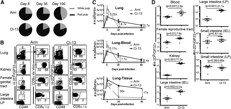
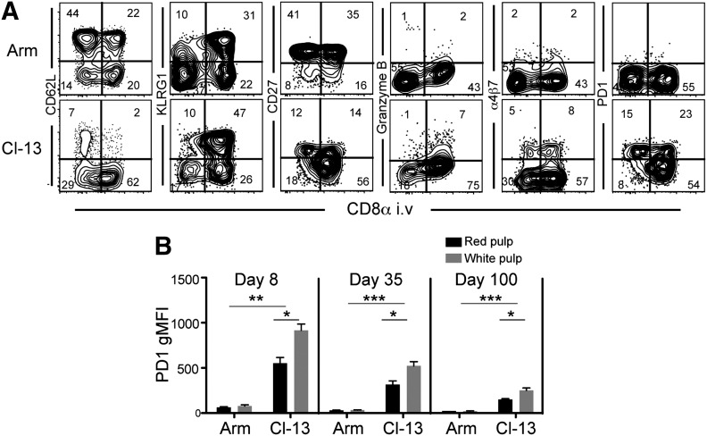
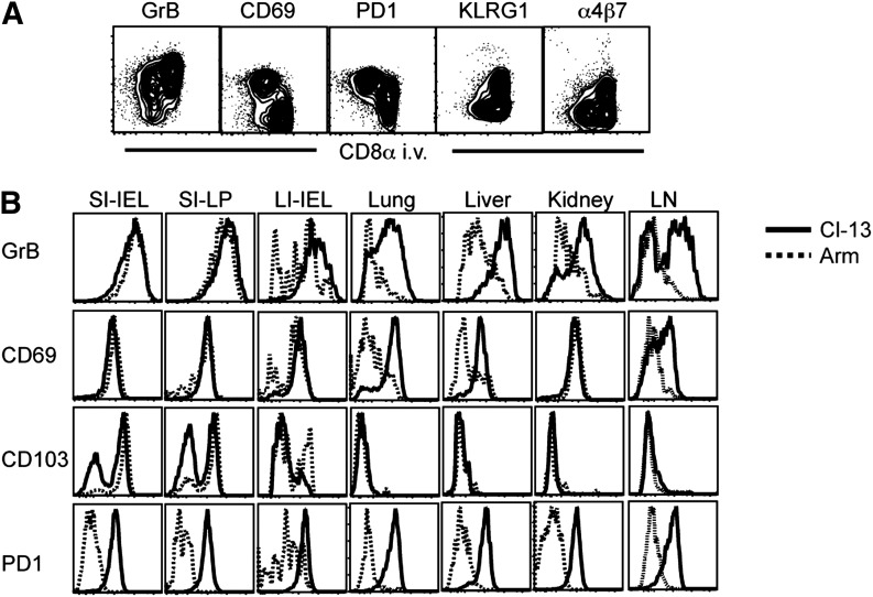
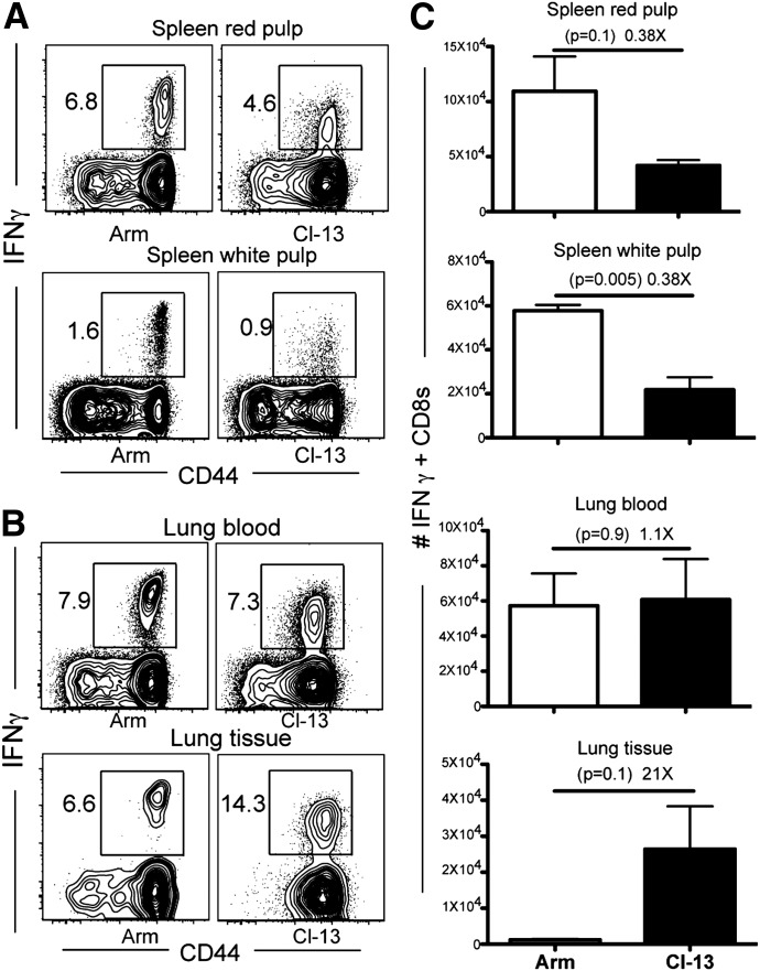
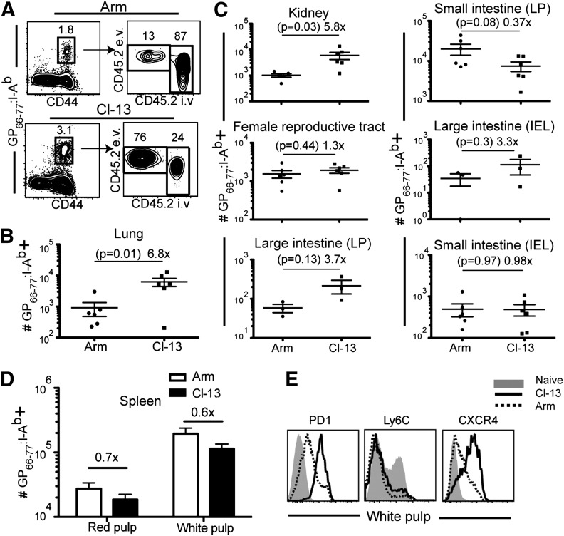
Vaccines are desired that maintain abundant memory T cells at nonlymphoid sites of microbial exposure, where they may be anatomically positioned for immediate pathogen interception. Here, we test the impact of antigen persistence on mouse CD8 and CD4 T cell distribution and differentiation by comparing responses to infections with different strains of LCMV that cause either acute or chronic infections. We used in vivo labeling techniques that discriminate between T cells present within tissues and abundant populations that fail to be removed from vascular compartments, despite perfusion. LCMV persistence caused up to ∼30-fold more virus-specific CD8 T cells to distribute to the lung compared with acute infection. Persistent infection also maintained mucosal-homing α4β7 integrin expression, higher granzyme B expression, alterations in the expression of the TRM markers CD69 and CD103, and greater accumulation of virus-specific CD8 T cells in the large intestine, liver, kidney, and female reproductive tract. Persistent infection also increased LCMV-specific CD4 T cell quantity in mucosal tissues and induced maintenance of CXCR4, an HIV coreceptor. This study clarifies the relationship between viral persistence and CD4 and CD8 T cell distribution and mucosal phenotype, indicating that chronic LCMV infection magnifies T cell migration to nonlymphoid tissues.

Keywords: CD8 T cells; homing.


Figures

Figure 1.
Figure 1.. Comparison of LCMV Armstrong and Cl-13 persistence in mouse tissues.
C57BL/6J mice were infected with LCMV Armstrong (Arm) or Cl-13, and on the days indicated, tissue sections were stained with DAPI (gray) and LCMV nucleoprotein-specific mAb (green) to assess viral replication. Images are representative of three independent experiments, with ≥3 mice/infection condition. Original scale bars = 100 μm.
Figure 2.
Figure 2.. LCMV persistence affects distribution of virus-specific CD8 T cells.
CD8 T cells were isolated from tissues on various days after LCMV Armstrong or Cl-13 infection. (A) H-2Db/GP33 MHC class I tetramer+ CD8 T cells were enumerated independently in the red and white pulp of the spleen via intravascular staining (see Materials and Methods). (B) The frequency and intravascular staining pattern of H-2Db/GP33-specific CD8 T cells in various tissues, 35 days after infection. Plots gated on CD8+ lymphocytes, representative of 10 mice from 3 independent experiments. LP, Lamina propria. (C) The kinetics of the H-2Db/GP33-specific CD8 T cell response in the entire lung and the compartments that were stained with (lung blood) or protected from (lung tissue) intravascular-injected antibody. (D) The number of H-2Db/GP33-specific CD8 T cells protected from intravascular CD8α staining in various tissues (including peripheral blood, where all CD8 T cells are stained with i.v.-injected antibody) was compared, 35 days after each infection. Error bars indicate sem.
Figure 3.
Figure 3.. Sublocalization within spleen and LCMV persistence delineates CD8 T cell phenotype.
C57BL/6J mice were infected with LCMV Armstrong or Cl-13. One hundred days later, mice were injected with anti-CD8α i.v., and splenocytes were isolated. (A) CD8α staining versus the indicated markers, gated on H-2Db/GP33-specific CD8 T cells. (B) Geometric mean fluorescence intensity (gMFI) of PD-1 expression on H-2Db/GP33-specific CD8 T cells isolated from red or white pulp. Data are representative of 9 mice from 3 independent experiments. Error bars indicate sem; ***P < 0.001; **P < 0.01; *P < 0.05.
Figure 4.
Figure 4.. LCMV persistence regulates effector differentiation and mucosal-homing molecule expression in nonlymphoid organs.
H-2Db/gp33 CD8 T cells were phenotyped by flow cytometry, 35 days after LCMV Armstrong or Cl-13 infection. Anatomic distribution was determined by permissiveness to intravascular anti-CD8α staining. (A) Antigen-specific CD8 T cells isolated from lung after Cl-13 infection. (B) H-2Db/GP33 MHC I tetramer+ cells that were protected from intravascular staining after LCMV Armstrong (dashed black histogram) or Cl-13 (solid black histogram) infection isolated from indicated tissues. Data are representative of 9 mice from 3 independent experiments. GrB, Granzyme B; SI, small intestine; LI, large intestine; LN, lymph node.
Figure 5.
Figure 5.. There are more IFN-γ-competent CD8 T cells in lung after persistent LCMV infection.
Forty-five days after LCMV Armstrong or Cl-13 infection, lymphocytes were isolated, stimulated with gp33 for 3 h in vitro, and then stained with an IFN-γ antibody. Representative flow cytometry plots showing the frequency of IFN-γ-positive cells on CD8+ lymphocytes isolated from (A) spleen and (B) lung blood and tissue. (C) As in A and B, however, IFN-γ+ cells in each location were enumerated. Error bars indicate sem.
Figure 6.
Figure 6.. Persistent LCMV drives enhanced expression of nonlymphoid-homing markers on CD4 T cells and preferential distribution to nonlymphoid tissues.
The phenotype of GP66–77:I-Ab MHC II tetramer+ LCMV-specific CD4 T cells from various tissues was analyzed, 35 days after LCMV Armstrong or Cl-13 infection. (A) Representative intravascular staining on MHC II tetramer+ CD4 T cells in lung. (B) Enumeration of isolated GP66–77:I-Ab MHC II tetramer+ CD4 T cells that were protected from intravascular staining in the lung, (C) indicated tissues, and (D) spleen white pulp (intravascular staining + red pulp cells are also shown). Error bars represent sem. (E) Comparison of PD-1, Ly6C, and CXCR4 expression on spleen white pulp-derived GP66–77:I-Ab MHC II tetramer+ CD4 T cells after LCMV Armstrong (dashed black line) or Cl-13 (solid black line) infection. Phenotype of naïve CD44lo splenocytes (solid gray histogram) is shown for comparison. Data are representative of two independent experiments totaling ≥6 mice/group.

Comment in

References

    1. Cauley L. S., Lefrançois L. (2013) Guarding the perimeter: protection of the mucosa by tissue-resident memory T cells. Mucosal Immunol. 6, 14–23. - PMC - PubMed
    1. Mueller S. N., Gebhardt T., Carbone F. R., Heath W. R. (2013) Memory T cell subsets, migration patterns, and tissue residence. Annu. Rev. Immunol. 31, 137–161. - PubMed
    1. Kohlmeier J. E., Woodland D. L. (2009) Immunity to respiratory viruses. Annu. Rev. Immunol. 27, 61–82. - PubMed
    1. Masopust D., Picker L. J. (2012) Hidden memories: frontline memory T cells and early pathogen interception. J. Immunol. 188, 5811–5817. - PMC - PubMed
    1. Gebhardt T., Wakim L. M., Eidsmo L., Reading P. C., Heath W. R., Carbone F. R. (2009) Memory T cells in nonlymphoid tissue that provide enhanced local immunity during infection with herpes simplex virus. Nat. Immunol. 10, 524–530. - PubMed

Publication types

MeSH terms