. 2014 Nov 13;55(12):8067-76.
doi: 10.1167/iovs.14-14722.

Shear stress-triggered nitric oxide release from Schlemm's canal cells

Affiliations

Shear stress-triggered nitric oxide release from Schlemm's canal cells

Nicole E Ashpole et al. Invest Ophthalmol Vis Sci. .

Abstract

Purpose: Endothelial nitric oxide (NO) synthase is regulated by shear stress. At elevated intraocular pressures when the Schlemm's canal (SC) begins to collapse, shear stress is comparable with that in large arteries. We investigated the relationship between NO production and shear stress in cultured human SC cells.

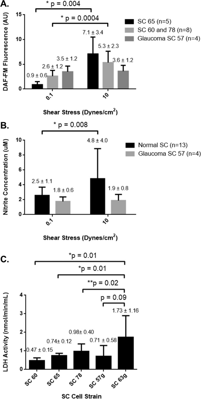
Methods: Schlemm's canal endothelial cells isolated from three normal and two glaucomatous human donors were seeded into Ibidi flow chambers at confluence, cultured for 7 days, and subjected to steady shear stress (0.1 or 10 dynes/cm(2)) for 6, 24, or 168 hours. Cell alignment with flow direction was monitored, and NO production was measured using 4-amino-5-methylamino-2',7'-difluorofluorescein (DAF-FM) and Griess reagents. Human trabecular meshwork (TM) and umbilical vein endothelial cells (HUVECs) were used as controls.

Results: Normal SC strains aligned with the direction of flow by 7 days. Comparing 0.1 vs. 10 dynes/cm(2), NO levels increased by 82% at 24 hours and 8-fold after 7 days by DAF-FM, and similar results were obtained with Griess reagent. Shear responses by SC cells at 24 hours were comparable with HUVECs, and greater than TM cells, which appeared shear-insensitive. Nitric oxide production by SC cells was detectable as early as 6 hours and was inhibited by 100 μM nitro-L-arginine methyl ester. Two glaucomatous SC cell strains were either unresponsive or lifted from the plate in the face of shear.

Conclusions: Shear stress triggers NO production in human SC cells, similar to other vascular endothelia. Increased shear stress and NO production during SC collapse at elevated intraocular pressures may in part mediate IOP homeostasis.

Keywords: aqueous humor; conventional outflow; eNOS; glaucoma; intraocular pressure.


Figures

Figure 1
Figure 1
Alignment of normal SC and HUVECs induced by shear stress. Phase-contrast images of HUVECs exposed to 0.1 dynes/cm2 or 10 dynes/cm2 for 24 hours show alignment at the higher value of shear stress. The direction of flow/shear is indicated by arrows. Similarly, SC cells were exposed to shear stress for 1 week and cell alignment was assessed. Images are representative, and show data from one experiment of five total for HUVECs and of eight total for SCs, using two SC cell strains.
Figure 2
Figure 2
Histograms showing cultured endothelial cell alignment relative to the direction of flow/shear (defined as 0°) when exposed to shear stresses of 0.1 or 10 dynes/cm2. (A) Displays pooled cell orientation data obtained from images of HUVECs (mean ± SD, n = 5) exposed to shear stress for 24 hours. The dashed line indicates the expected frequency of 16.7% for each 15° bin, corresponding to the case of uniform distribution between bins (random cell orientation). (B) Shows distribution of Schlemm's canal cells across bins (combined data from two normal strains, mean ± SD, n = 8) exposed to shear stresses for 1 week. Significant differences were determined by comparing the sample numbers in each individual bin at each shear stress level to the expected frequency. *P < 0.05.
Figure 3
Figure 3
Nitric oxide levels assessed by DAF-FM fluorescence in HUVECs and Schlemm's canal (SC) cells exposed to shear stress. We exposed HUVECs and SC cells to 0.1 dynes/cm2 or 10 dynes/cm2 and DAF-FM fluorescence was evaluated using fluorescence microscopy. We exposed HUVECs to shear for 24 hours while SC cells were exposed to shear for 1 week. At the end of each experiment, a DAF-FM probe was applied. Shown are images of a single field of view (five total taken) from a single condition, representative of five total experiments for HUVECs and eight total experiments using two SC cell strains.
Figure 4
Figure 4
Nitric oxide production by HUVECs exposed to shear stress for 24 hours. Cells were exposed to shears of 0.1 or 10 dynes/cm2. (A) Shows the quantification of NO content from DAF-FM fluorescence and (B) nitrite concentration from Griess reagent analysis (mean ± SD, n = 5).
Figure 5
Figure 5
Nitric oxide production by normal Schlemm's canal cells exposed to shear stress for 1 week. Cells were exposed to 0.1 or 10 dynes/cm2 for 1 week. (A) Shows the quantification of NO content derived from DAF-FM fluorescence and (B) quantification of nitrite concentration from Griess reagent analysis for normal SC cell strains. Shown are combined data from two normal cell strains (SC60 and 65, mean ± SD, n = 8).
Figure 6
Figure 6
Nitric oxide production and LDH release by Schlemm's canal cells exposed to shear stress for 24 hours. Cells were exposed to 0.1 or 10 dynes/cm2 for 24 hours and NO content was measured by (A) DAF-FM fluorescence and (B) Griess reagent for normal SC cell strains (combined data from three normal strains) and one glaucomatous cell strain (SC57g). (C) LDH activity assay to estimate cell viability in cells exposed to 10 dynes/cm2 of shear for 24 hours. All data shown are expressed as mean values ± SD.
Figure 7
Figure 7
Nitric oxide production in Schlemm's canal cells and HUVECs exposed to 10 dynes/cm2 shear stress ± L-NAME for 6 hours. (A) Shows the quantification of NO content from DAF-FM fluorescence and (B) nitrite concentration from Griess reagent analysis (mean ± SD).
Figure 8
Figure 8
Nitric oxide production by trabecular meshwork cells exposed to shear stress for 24 hours. Shown are combined data (mean ± SD) from two cell strains exposed to 0.1 or 10 dynes/cm2 for 24 hours. NO content was measured by (A) DAF-FM fluorescence and (B) Griess reagent analysis.

References

    1. Predescu D, Predescu S, Shimizu J, Miyawaki-Shimizu K, Malik AB. Constitutive eNOS-derived nitric oxide is a determinant of endothelial junctional integrity. Am J Physiol Lung Cell Mol Physiol. 2005; 289: L371–L381. - PubMed
    1. Colasanti M, Suzuki H. The dual personality of NO. Trends Pharmacol Sci. 2000; 21: 249–252. - PubMed
    1. Harrison DG. Cellular and molecular mechanisms of endothelial cell dysfunction. J Clin Invest. 1997; 100: 2153–2157. - PMC - PubMed
    1. Harrison DG, Cai H. Endothelial control of vasomotion and nitric oxide production. Cardiol Clin. 2003; 21: 289–302. - PubMed
    1. Becquet F, Courtois Y, Goureau O. Nitric oxide in the eye: Multifaceted roles and diverse outcomes. Surv Ophthamol. 1997; 42: 71–82. - PubMed

Publication types

MeSH terms