. 2015 Jan 15;128(2):364-72.
doi: 10.1242/jcs.161323. Epub 2014 Nov 13.

INPP5E interacts with AURKA, linking phosphoinositide signaling to primary cilium stability

Affiliations

INPP5E interacts with AURKA, linking phosphoinositide signaling to primary cilium stability

Olga V Plotnikova et al. J Cell Sci. .

Abstract

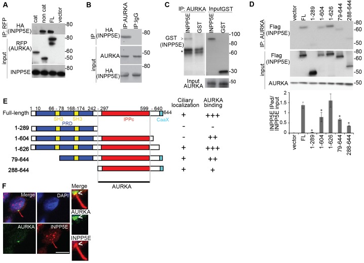
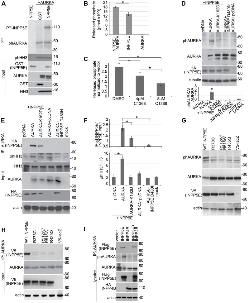
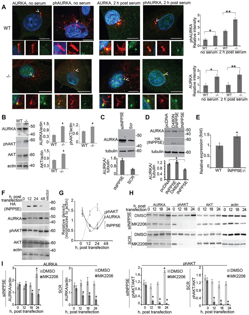
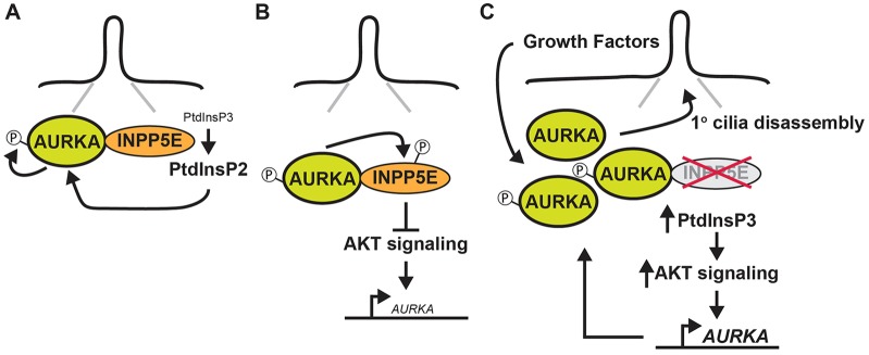
Mutations in inositol polyphosphate 5-phosphatase E (INPP5E) cause the ciliopathies known as Joubert and MORM syndromes; however, the role of INPP5E in ciliary biology is not well understood. Here, we describe an interaction between INPP5E and AURKA, a centrosomal kinase that regulates mitosis and ciliary disassembly, and we show that this interaction is important for the stability of primary cilia. Furthermore, AURKA phosphorylates INPP5E and thereby increases its 5-phosphatase activity, which in turn promotes transcriptional downregulation of AURKA, partly through an AKT-dependent mechanism. These findings establish the first direct link between AURKA and phosphoinositide signaling and suggest that the function of INPP5E in cilia is at least partly mediated by its interactions with AURKA.

Keywords: AURKA; INPP5E; Primary cilium.


Figures

Fig. 1.
Fig. 1.
AURKA directly interacts with INPP5E. (A) RFP-fused full-length (FL) AURKA and catalytic (cat; amino acids 132–403) and non-catalytic (non cat; amino acids 1–131) domains were coexpressed with HA–INPP5E in HEK293 cells and immunoprecipitated (IP) with anti-RFP antibody. Immunoprecipitates were western blotted with the antibodies indicated. (B) Endogenous AURKA was immunoprecipitated from IMCD3 cells expressing HA–INPP5E and blotted as indicated. Immunoglobulin G (IgG) was used as negative control for immunoprecipitation. (C) AURKA was immunoprecipitated from the in vitro mixture containing GST–INPP5E or GST only and recombinant His–AURKA, and immunoprecipitates were probed by western blotting (anti-GST, left panel). Starting material is shown on the right (Input). The asterisk indicates the heavy chain of IgG. (D) Anti-AURKA antibody was used to immunoprecipitate endogenous AURKA from whole IMCD3 cell lysates expressing vector alone, FLAG-tagged full-length INPP5E and INPP5E deletion mutants, followed by western blotting as indicated. Molecular masses are indicated in kDa. The graph indicates the ratio of precipitated to total INPP5E, quantified from analysis of blots from three independent experiments. Data show the mean±s.e.m.; *P<0.001 compared to full-length INPP5E (Student's t-test). (E) Schematic representation of INPP5E variants used for AURKA binding studies. The proline-rich domain (PRD) is indicated in blue, inositol polyphosphate phosphatase catalytic (IPPc) domain in red, CaaX motif is in light blue and the SH3 domain is in yellow. The extent to which each construct localizes to the cilium and binds to AURKA is indicated on the right. +, weak binding; ++, medium binding; +++, strong binding; −, absence of binding. (F) Immunofluorescent detection of AURKA (green) and INPP5E (red) in IMCD3 cells demonstrating basal body localization of AURKA and ciliary axoneme localization of INPP5E, overlapping with AURKA signal at basal end of cilium (arrowheads). 4′,6-diamidino-2-phenylindole (DAPI) staining indicates the nucleus (blue). Scale bar: 10 µm. Insets show a magnification of the basal body and cilium.
Fig. 2.
Fig. 2.
Interactions between AURKA and INPP5E affect the activity of both proteins. (A) In vitro kinase assay. The upper panel indicates γ-[32P]ATP signal corresponding to phosphorylated GST–INPP5E (∼110 kDa), auto-phosphorylated (ph)AURKA (∼50 kDa) and phosphorylated histone H3 (phHH3, ∼16 kDa). The lower panel shows input proteins western blotted as indicated. (B) Recombinant active AURKA activates the hydrolysis of PtdIns(3,4,5)P3 by INPP5E (n = 3 for each sample). Data show the mean±s.e.m.; *P = 0.014 (Student's t-test). (C) Quantification of INPP5E 5-phosphatase activity against PtdIns(3,4,5)P3 upon AURKA inhibition with the indicated concentrations of C1368 in HEK293 cells overexpressing HA–INPP5E or empty vector. Data show the mean±s.e.m.; *P<0.01 (Student's t-test). Results were normalized to those of the vector-expressing cells to reduce background caused by the presence of other phosphatases in the cells. Non-normalized data are shown in supplementary material Fig. S2D. (D) HEK293 cells were co-transfected with plasmids expressing the indicated proteins. Cell lysates were separated and probed with the antibodies indicated. Blots from three independent experiments were quantified and the relative phosphorylation of AURKA was calculated (lower panel). Data show the mean±s.e.m.; *P = 0.002 (Student's t-test). (E) HEK293 cells were co-transfected with the indicated plasmids. AURKA was immunoprecipitated (IP) and a fraction of the sample was used in an in vitro kinase assay to detect AURKA auto-phosphorylation and AURKA activity towards HH3. A second fraction was used to assess AURKA immunoprecipitation with HA–INPP5E. The kinase dead AURKA mutant K162D was used as a negative control for specificity of AURKA phosphorylation. (F) Graphs indicate the ratio of phosphorylated HH3 to total HH3 (upper panel) and of immunoprecipitated INPP5E to input in cell lysates. Data were quantified from three blots and show the mean±s.e.m.; *P<0.05 (Student's t-test). (G,H) Western blot analysis of lysates (G) and immunoprecipitates (H) from HEK293 cells co-transfected with RFP–AURKA and either wild-type (WT) INPP5E or the indicated INPP5E JBTS mutants. Plasmid V5-LacZ was used as a negative control for immunoprecipitation. (I) Western blot analysis of precipitates and lysates from HEK293 cells co-transfected with RFP–AURKA and the indicated phosphatases. Molecular masses are indicated in kDa.
Fig. 3.
Fig. 3.
AURKA levels are regulated by INPP5E. (A) Immunofluorescence detection of AURKA (green) or T288-phosphorylated AURKA (phAURKA, green), acetylated α-tubulin (cilia, red) and DAPI (DNA, blue) in primary fibroblasts from Inpp5e+/+ (WT) and Inpp5e−/− (−/−) embryos with or without serum as indicated. Arrowheads indicate primary cilia. Scale bar: 10 µm. Insets show magnifications of the centrosome and cilium. The graphs show the relative intensity of phosphorylated (ph)AURKA or AURKA in the basal body from three triplicate assays measuring 50 cells per assay. Data show the mean±s.e.m.; *P = 0.003, **P = 0.013 (for phosphorylated AURKA) and *P = 0.0012, **P = 0.014 (for AURKA; Student's t-test). (B) Western blot analysis of Ser473-phosphorylated AKT and AURKA in Inpp5e+/+ and Inpp5e−/− embryonic fibroblasts. Graphs indicate the ratio of either AURKA, phosphorylated AKT or AKT to actin loading control, from analysis of three blots. Data show the mean±s.e.m.; *P<0.05. (C) IMCD3 cells were transfected with INPP5E-specific siRNA (siINPP5E) or scrambled control siRNA (Scr). Graphs indicate the ratio of AURKA to tubulin loading control, quantified from three blots. Data show the mean±s.e.m.; *P = 0.0045. (D) IMCD3 cells were transfected and western blotted as indicated. Graphs indicate the ratio of AURKA to tubulin loading control, quantified from three blots; Data show the mean±s.e.m.; *P≤0.0032 (compared with pcDNA). (E) mRNA expression levels of Aurka were determined from Inpp5e+/+ and Inpp5e−/− embryonic fibroblast cultures by real-time RT-PCR, and data are presented as the fold change relative to the wild-type control. Data show the mean±s.e.m.; *P = 0.017. (F) Plasmids expressing HA–INPP5E were transfected into HEK293 cells, and lysates were collected at the indicated time-points. Western blotting was performed with the indicated antibodies. Molecular masses are indicated in kDa. (G) AURKA levels and the relative phosphorylation of AKT and INPP5E were quantified from the results of the experiment presented in F. Data are expressed as the mean±s.e.m. (three experiments). (H) IMCD3 cells were siRNA transfected then treated for 12 h with 10 µM AKT inhibitor MK2206 or DMSO. Lysates were collected at the indicated time-points and western blotted as indicated. (I) The ratio of AURKA or phosphorylated AKT to loading control actin under different conditions following AUKRA inhibition, as quantified from analysis of three blots. Data show the mean±s.e.m.; *P<0.01.
Fig. 4.
Fig. 4.
Functional role of the interaction between AURKA and INPP5E. (A) Cilia in Inpp5e+/+ (WT) and Inpp5e−/− (−/−) primary mouse embryonic fibroblasts (72 h under low-serum conditions) were induced to disassemble in the presence of 2 µM AURKA inhibitor (C1368) or DMSO control. All images show merged panels of the ciliary marker acetylated α-tubulin (green), the centrosome and basal body marker γ-tubulin (red) and DAPI (blue). Arrowheads indicate primary cilia. Scale bars: 10 µm. The percentage of ciliated cells at times before and after the indicated treatments are indicated in the graph on the right. Three triplicate assays were undertaken, counting 100 cells per assay. Data show the mean±s.e.m.; *P<0.01; ns, not significant (Student's t-test). (B) Immunofluorescence images of siRNA-treated (siINPP5E) IMCD3 cells with 2 µM C1368 AURKA inhibitor or DMSO control, grown in 3D Matrigel culture. Scr, scrambled control RNA; AcTub, acetylated tubulin (green, cilia); E-cadh, E-cadherin (red, cell membrane); DAPI, DNA staining (blue). (C) Quantitative analysis of spheroid numbers per well. Data show the mean±s.e.m.; *P<0.01 (Student's t-test).
Fig. 5.
Fig. 5.
A model for reciprocal interactions between INPP5E and AURKA. (A) At the primary cilium we propose that hydrolysis of PtdIns(3,4,5)P3 by INPP5E alters the levels of different phosphoinositide species and, in so doing, mediates the functional interaction between INPP5E and AURKA, resulting in the activation of the latter protein through auto-phosphorylation. (B) Activated AURKA binds to and phosphorylates INPP5E, increasing its phosphatase activity and reducing AURKA transcription (small font) downstream of AKT. (C) When INPP5E is deleted or mutated, increased AKT signaling elevates AURKA transcription (large font), resulting in a generalized accumulation of AURKA protein at the cilia. In response to physiological stimuli, we propose that the increased pool of AURKA at this site drives the abnormal cilia disassembly phenotype observed in cells lacking INPP5E.

References

    1. Bertotti A., Burbridge M. F., Gastaldi S., Galimi F., Torti D., Medico E., Giordano S., Corso S., Rolland-Valognes G., Lockhart B. P. <et al.(2009). Only a subset of Met-activated pathways are required to sustain oncogene addiction. Sci. Signal. 2, ra80 10.1126/scisignal.2000643 - DOI - PubMed
    1. Bielas S. L., Silhavy J. L., Brancati F., Kisseleva M. V., Al-Gazali L., Sztriha L., Bayoumi R. A., Zaki M. S., Abdel-Aleem A., Rosti R. O. <et al.(2009). Mutations in INPP5E, encoding inositol polyphosphate-5-phosphatase E, link phosphatidyl inositol signaling to the ciliopathies. Nat. Genet. 41, 1032–1036 10.1038/ng.423 - DOI - PMC - PubMed
    1. Cheetham G. M., Knegtel R. M., Coll J. T., Renwick S. B., Swenson L., Weber P., Lippke J. A., Austen D. A. (2002). Crystal structure of aurora-2, an oncogenic serine/threonine kinase. J. Biol. Chem. 277, 42419–42422 10.1074/jbc.C200426200 - DOI - PubMed
    1. Conduit S. E., Dyson J. M., Mitchell C. A. (2012). Inositol polyphosphate 5-phosphatases; new players in the regulation of cilia and ciliopathies. FEBS Lett. 586, 2846–2857 10.1016/j.febslet.2012.07.037 - DOI - PubMed
    1. Gautschi O., Heighway J., Mack P. C., Purnell P. R., Lara P. N., Jr and Gandara D. R. (2008). Aurora kinases as anticancer drug targets. Clin. Cancer Res. 14, 1639–1648 10.1158/1078-0432.CCR-07-2179 - DOI - PubMed

Publication types

MeSH terms

Supplementary concepts