. 2014 Aug 6;24(29):4615-4624.
doi: 10.1002/adfm.201400526.

Highly tunable elastomeric silk biomaterials

Affiliations

Highly tunable elastomeric silk biomaterials

Benjamin P Partlow et al. Adv Funct Mater. .

Abstract

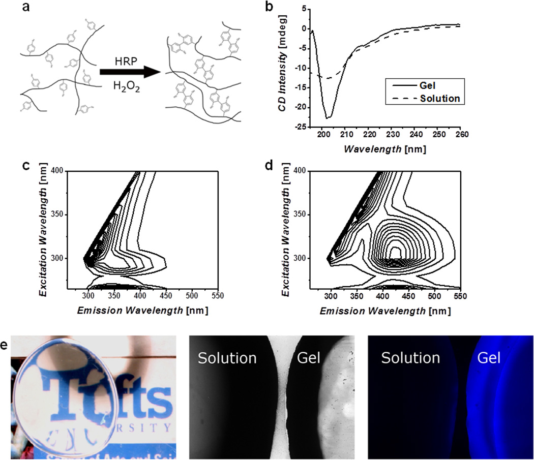
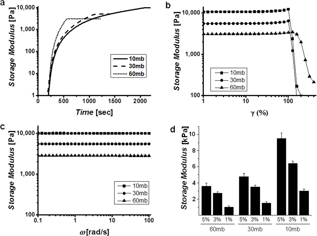
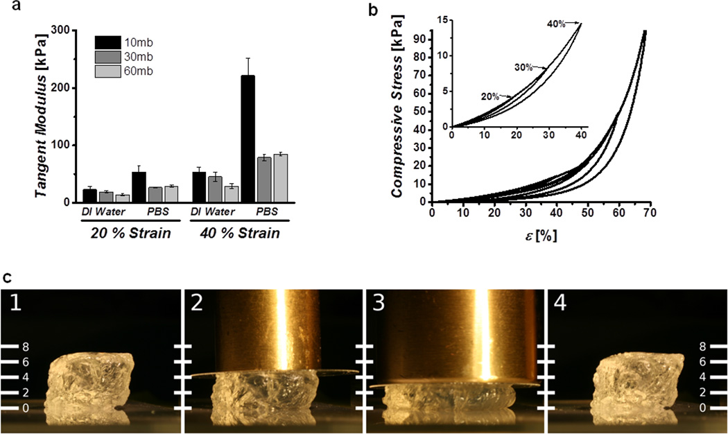
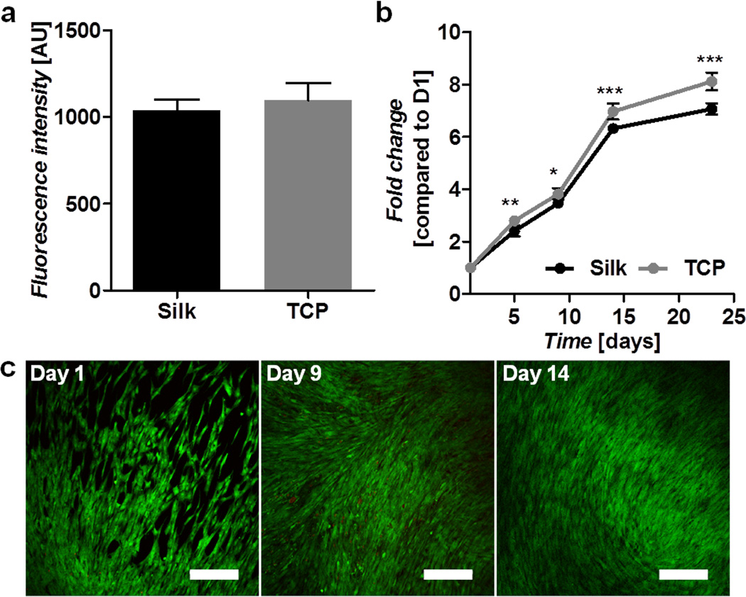
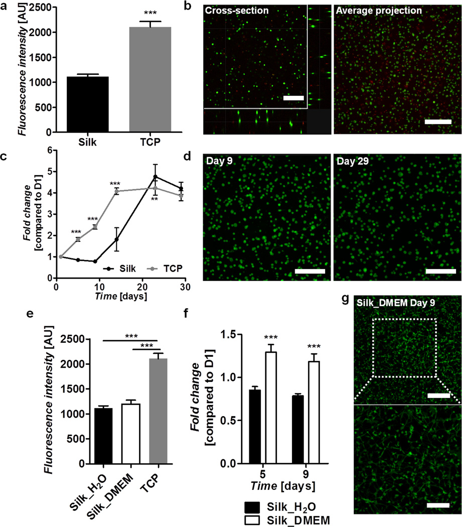
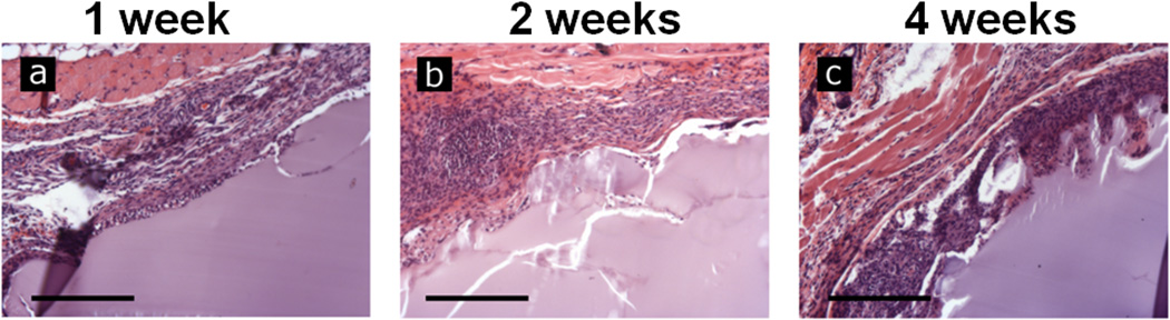
Elastomeric, fully degradable and biocompatible biomaterials are rare, with current options presenting significant limitations in terms of ease of functionalization and tunable mechanical and degradation properties. We report a new method for covalently crosslinking tyrosine residues in silk proteins, via horseradish peroxidase and hydrogen peroxide, to generate highly elastic hydrogels with tunable properties. The tunable mechanical properties, gelation kinetics and swelling properties of these new protein polymers, in addition to their ability to withstand shear strains on the order of 100%, compressive strains greater than 70% and display stiffness between 200 - 10,000 Pa, covering a significant portion of the properties of native soft tissues. Molecular weight and solvent composition allowed control of material mechanical properties over several orders of magnitude while maintaining high resilience and resistance to fatigue. Encapsulation of human bone marrow derived mesenchymal stem cells (hMSC) showed long term survival and exhibited cell-matrix interactions reflective of both silk concentration and gelation conditions. Further biocompatibility of these materials were demonstrated with in vivo evaluation. These new protein-based elastomeric and degradable hydrogels represent an exciting new biomaterials option, with a unique combination of properties, for tissue engineering and regenerative medicine.

Keywords: biomaterials; biopolymers; elastomers; hydrogels; silk.


Figures

Figure 1
Figure 1. Chemistry and structural characterization
a) Schematic representation of the crosslinking of tyrosine residues on silk molecules, these covalent bonds allow for chain extension creating highly elastic hydrogels. b) Circular Dichroism (CD) spectra of silk solution and enzymatically formed hydrogels, show a change to a helical structure and not β-sheet as found in other silk materials. Fluorescence excitation-emission spectra of solution (c) and gel (d) confirm the formation of dityrosine bonds. e) The resultant hydrogels are optically clear and exhibit a blue fluorescence when irradiated with UV that is not present in the precursor solution.
Figure 2
Figure 2. Rheological properties of hydrogels
Representative gelation kinetics (a), strain sweeps (b) and frequency sweeps (c) of different molecular weight gels show controllable kinetics and final mechanical properties in highly resilient, frequency independent gels. Curves indicated were performed at a protein concentration of 5% w/v in DI water. d) Final storage modulus as a function of molecular weight and concentration, demonstrate the wide range of stiffness achievable.
Figure 3
Figure 3. Compressive properties of hydrogels
a) Tangent moduli of 5% w/v cast gels swelled in water and PBS show strain and molecular weight dependence. b) Cyclic compression curves of gels immersed in PBS showing excellent recovery below 70% strain, inset highlights complete recovery below 40% strain. c) Image of hydrogel undergoing ~50% compression, under 50 g (2) and 100 g (3) brass weights and showing complete recovery after removal (4). Scale units are in milimeters.
Figure 4
Figure 4. Human mesenchymal stem cell interactions on silk gel surfaces
a) Cell attachment on silk and TCP at day 1 post-seeding as determined by Alamar blue. b) Cell proliferation on silk gels and TCP over a 24-day period as determined by Alamar blue and presented as fold change compared to day 1. c) Live (green) and dead (red) cell staining on silk gels over a 14-day period. Scale bars are 300 µm.
Figure 5
Figure 5. Human mesenchymal stem cells encapsulated in silk gels
Silk gels were formed in water (a–d) or DMEM (e–g). a) Cell survival following encapsulation in silk gels compared to cells seeded on TCP as determined by Alamar blue at day 1 post-encapsulation. b) Live (green) and dead (red) staining on cells encapsulated in silk gels at day 1 post-encapsulation showing a cross-section and an average projection of a 352 µm thick image stack. c) Cell proliferation on silk gels and TCP over a 29 day period as determined by Alamar blue and presented as fold change compared to day 1. d) Live (green) and dead (red) staining of cells encapsulated in silk gels at days 9 and 29 post-seeding showing an average projection of 212 µm and 212 µm thick image stacks respectively. Scale bars are 300 µm. e) Cell survival following encapsulation in silk gels formed in water and DMEM compared to cells seeded on TCP as determined by Alamar blue at day 1 post-encapsulation. f) Comparison of cell proliferation in silk gels formed in water and DMEM as determined by Alamar blue at days 5 and 9 post-encapsulation and presented as fold change compared to day 1. g) Live (green) and dead (red) staining on cells encapsulated in a silk gel formed in DMEM at day 9 post-encapsulation showing an average projection of a 180 µm thick mage stack. Scale bars are 300 µm (top) and 175 µm (bottom).
Figure 6
Figure 6. In vivo interactions with implanted silk gels
Preformed hydrogels of 6% w/v were implanted subcutaneously in a mouse model. Gels were explanted and examined histologically by H&E staining 1 (a), 2 (b) and 4 (c) weeks following implantation. The hydrogels showed progressive cell infiltration and degradation as the duration was increased. Scale bars are 250 µm.

References

    1. Seliktar D. Science. 2012;336:1124. - PubMed
    1. Engler AJ, Sen S, Sweeney HL, Discher DE. Cell. 2006;126:677. - PubMed
    1. Hoffman AS. Advanced Drug Delivery Reviews. 2012
    2. Peppas N, Huang Y, Torres-Lugo M, Ward J, Zhang J. Annual Review of Biomedical Engineering. 2000;2:9. - PubMed
    1. Rai R, Tallawi M, Grigore A, Boccaccini AR. Progress in polymer science. 2012;37:1051.
    1. Zdrahala RJ, Zdrahala IJ. Journal of biomaterials applications. 1999;14:67. - PubMed