. 2014 Nov 12:3:585.
doi: 10.1038/bonekey.2014.80. eCollection 2014.

Generation of rodent and human osteoblasts

Affiliations

Generation of rodent and human osteoblasts

Sarah E B Taylor et al. Bonekey Rep. .

Abstract

This paper describes the isolation, culture and staining of primary osteoblasts from neonatal rodents and human samples. The calvaria and long-bone assays allow direct measurement of bone matrix deposition and mineralisation, as well as producing osteoblasts at defined stages of differentiation for molecular and histological analysis. Culture of human osteoblasts enables cell function to be investigated in targeted patient groups. The described methods will provide a step-by-step guide of what to expect at each stage of the culture and highlight the varied tissue culture conditions required to successfully grow osteoblasts from different sources. A special focus of this paper is the methods used for analysis of bone mineralisation and how to ensure that nonspecific mineral deposition or staining is not quantified.


Conflict of interest statement

The authors declare no conflict of interest.

Figures
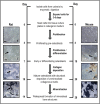
Figure 1
Figure 1
Calvarial osteoblast cultures. Rat calvarial osteoblast cultures are ∼14 days in duration when grown in DMEM. Representative images of unstained cell layers show that by day 4 of culture, a confluent monolayer of preosteoblasts is evident. At day 7, the cells are more compacted and organic matrix is starting to be deposited (as shown by the arrow). By day 10, there is abundant deposition of unmineralised collagenous matrix, and after 14 days there is widespread formation of mineralised bone structures. Mouse osteoblasts take ∼28 days to form mineralised matrix nodules. Representative images of mouse cells show the defined stages of differentiation at 4, 10, 20 and 28 days of culture. Scale bars: day 4/7=50 μm, days 10/14=500 μm.
Figure 2
Figure 2
Long-bone osteoblast cultures. Rat long-bone osteoblast cultures are ∼15–20 days in duration when grown in DMEM. Representative images of unstained cell layers and bone formation show that by day 5 of culture, a confluent monolayer of preosteoblasts is evident. At day 10, the cells are more compacted and the initial stages of collagen matrix deposition are evident. The collagen matrix is much denser by day 15 and mineralisation of collagenous structures can be seen. By day 20, there is abundant deposition of mineralised bone structures. Scale bars: whole-well scan=50 mm, phase contrast images=100 μm.
Figure 3
Figure 3
TEM images of the extracellular matrix produced by long-bone osteoblasts. Long-bone osteoblasts were grown in osDMEM for 28 days and then viewed by TEM. C=collagen fibres, which can be seen longitudinally and in transverse section (where they appear as dots). M=specific mineralisation of the collagenous extracellular matrix. Magnification at × 20 k and × 30 k scale bar=1 μm, × 60 k scale bar=500 nm.
Figure 4
Figure 4
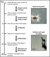
Human humeral head trabecular bone explant cultures. Human osteoblasts take ∼6–8 weeks to reach subconfluence when cultured in DMEM by the explant method. Representative images show unstained cell layers from explant bone at initial out growth and confluent monolayers of human humeral head trabecular osteoblasts before subculture. Following subculture mineralised bone structures are usually evident after ∼54 days. Scale bar=50 μm.
Figure 5
Figure 5
TNAP activity and soluble collagen levels in rat osteoblast cultures. TNAP activity and soluble collagen levels were measured at regular intervals throughout the osteoblast culture period. (a) TNAP activity was 2.5-fold and ⩽fourfold higher in differentiating and mature osteoblasts, respectively, compared with proliferating, preosteoblasts. (b) Soluble collagen levels were increased up to twofold in mature osteoblasts. Values are mean±s.e.m.; **P<0.01, ***P<0.001.
Figure 6
Figure 6
TNAP activity and mineralisation in human trabecular osteoblasts from normal and osteoarthritic patients. The level of mineralised nodule formation increases with culture time in human osteoblasts from (a) normal (30 years old) and (b) osteoarthritic patients (86 years old). (c) Representative whole-well scans of alizarin red stained human osteoblasts at different stages of culture. Human cells appear to be unable to form the discrete trabecular bone structures characteristic of neonatal rodent osteoblasts; instead, the mineralisation is more widespread. Human osteoblasts from (d) normal and (e) osteoarthritic patients display TNAP activity throughout the culture period. (f) Representative images showing increased levels of TNAP staining with osteoblast differentiation. Values are mean±s.e.m.; **P<0.01, ***P<0.001. Scale bar=50 mm.
Figure 7
Figure 7
Different staining methods in primary calvarial osteoblast cultures. Images show 14-day rat and 28-day mouse osteoblast cultures either left unstained, or stained with TNAP, alizarin red or sirius red. The individual bone structures formed by rodent osteoblasts displayed marked differences in appearance. Rat osteoblasts typically produced smaller bone nodules that had a ‘trabecular-shaped' appearance, whereas in mouse osteoblast cultures the bone structures were larger but fewer in numbers. TNAP staining was closely associated with the mineralised matrix in rat osteoblast cultures but more widespread in mouse osteoblasts. Sirius red staining shows the presence of collagen fibres in these cultures. Scale bars=50 mm (whole-well scans) and 500 μm (phase contrast microscopy images).
Figure 8
Figure 8
Different staining methods in primary long-bone osteoblast cultures. Images show 20-day rat long-bone osteoblast cultures either left unstained, or stained with alizarin red or Masson's trichrome. The rat long-bone osteoblasts produce ‘trabecular-shaped' bony structures that display positive staining for calcium and collagen deposition. Scale bars=50 mm (whole-well scans) and 500 μm (phase contrast microscopy images).
Figure 9
Figure 9
Quantification of bone mineralisation by image analysis. The representative images in (a) are of the same cell layers before and after alizarin red staining; the stained images are examples of cultures that have been inadequately washed, leading to bleeding of the alizarin red into the surrounding cells (shown by the arrows). (b) Binary images of the same cell layers highlight the differences in the level of bone mineral detected between stained and unstained plates. (c) Comparison of stained and unstained plates by automated analysis shows that the level of bone formation is ⩽fourfold higher in the stained plates. Culture with excess β-glycerophosphate (10 mM) amplifies this problem due to the widespread nonspecific deposition of mineral. Scale bars=50 mm (whole-well scans) and 5 mm (nodule images). Values are mean±s.e.m.; ***P<0.001.

References

    1. Orriss IR, Taylor SE, Arnett TR. Rat osteoblast cultures. Methods Mol Biol 2012;816:31–41. - PubMed
    1. Dillon JP, Waring-Green VJ, Taylor AM, Wilson PJ, Birch M, Gartland A et al. Primary human osteoblast cultures. Methods Mol Biol 2012;816:3–18. - PubMed
    1. Bakker AD, Klein-Nulend J. Osteoblast isolation from murine calvaria and long bones. Methods Mol Biol 2012;816:19–29. - PubMed
    1. Taylor SEB, Key ML, Lander M, Orriss IR, Patel JJ, Arnett TR. A novel method for the isolation and culture of rat long bone osteoblasts. Bone 2009;44:S318–S319.
    1. Orriss IR, Utting JC, Brandao-Burch A, Colston K, Grubb BR, Burnstock G et al. Extracellular nucleotides block bone mineralization in vitro: evidence for dual inhibitory mechanisms involving both P2Y2 receptors and pyrophosphate. Endocrinology 2007;148:4208–4216. - PubMed

LinkOut - more resources