

Multiparametric evaluation of breast lesions using PET-MRI: initial results and future perspectives
- PMID: 25396329
- PMCID: PMC4616313
- DOI: 10.1097/MD.0000000000000115
Multiparametric evaluation of breast lesions using PET-MRI: initial results and future perspectives
Abstract
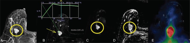
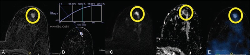
The purpose of this study was to evaluate the diagnostic accuracy of multiparametric evaluation of breast lesions combining information of dynamic contrast-enhanced magnetic resonance imaging (DCE-MRI), diffusion-weighted imaging (DWI), and F-fluoro-deoxi-glucose (F-FDG) positron emission tomography/computed tomography (PET-CT). After approval of the institutional research ethics committee, 31 patients with suspicious breast lesions on MRI performed F-FDG PET-CT with a specific protocol for breast evaluation. Patients' mean age was 47.8 years (range, 29-77 years). Positron emission tomography and magnetic resonance imaging (PET-MRI) images were fused. A lesion was considered positive on multiparametric evaluation if at least 1 of the following was present: washout/type 3 kinetic curve on DCE-MRI, restricted diffusion on DWI with minimum apparent diffusion coefficient value <1.00 × 10 mm/s, and abnormal metabolism on F-FDG PET-CT (higher than the physiologic uptake of the normal breast parenchyma). Thirty-eight lesions with histologic correlation were evaluated on the 31 included patients, being 32 mass lesions (84.2%), and 6 nonmass lesions (15.8%). Lesions' mean diameter was 31.1 mm (range, 8-94 mm). Multiparametric evaluation provided 100% sensitivity, 55.5% specificity, 87.9% positive predictive value, 100% negative predictive value, and 89.5% accuracy, with 29 true-positives results, 5 true-negatives, 4 false-positives, and no false-negative results. Multiparametric evaluation with PET-MRI functional data showed good diagnostic accuracy to differentiate benign from malignant breast lesions, reducing the number of unnecessary biopsies, without missing any diagnosis of cancer in our case series.
Conflict of interest statement
The authors have no funding and conflicts of interest to disclose.
Figures
References
-
- Fowler. AM. A molecular approach to breast imaging. J Nucl Med. 2014;55:177–180. - PubMed
-
- Peters NHGM, Borel Rinkes IHM, Zuithoff NPA, et al. Meta-analysis of MR imaging in the diagnosis of breast lesions. Radiology. 2008;246:116–124. - PubMed
-
- Brandão AC, Lehman CD, Partridge SC. Breast magnetic resonance imaging: diffusion-weighted imaging. Magn Reson Imaging Clin N Am. 2013;21:321–336. - PubMed
MeSH terms
Substances
LinkOut - more resources
Full Text Sources
Other Literature Sources
Medical
