

How does the structure of extraocular muscles and their nerves affect their function?
- PMID: 25397785
- PMCID: PMC4330282
- DOI: 10.1038/eye.2014.269
How does the structure of extraocular muscles and their nerves affect their function?
Abstract
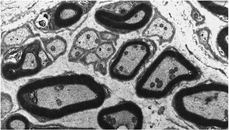
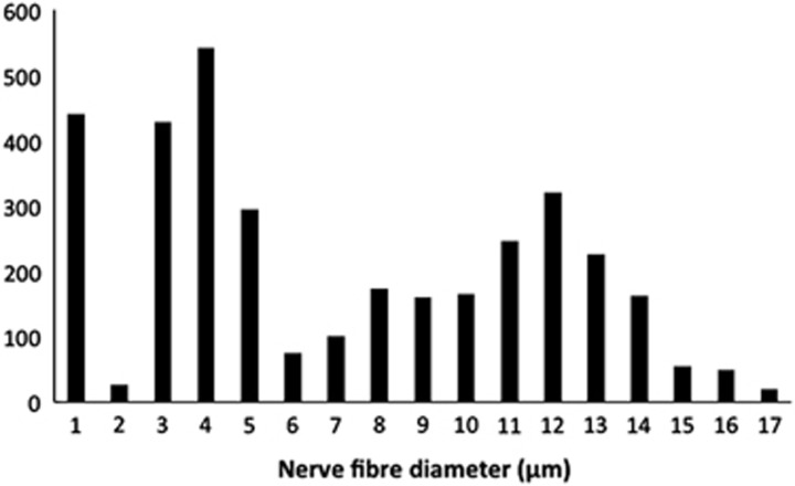
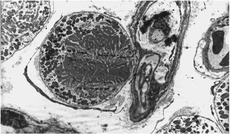
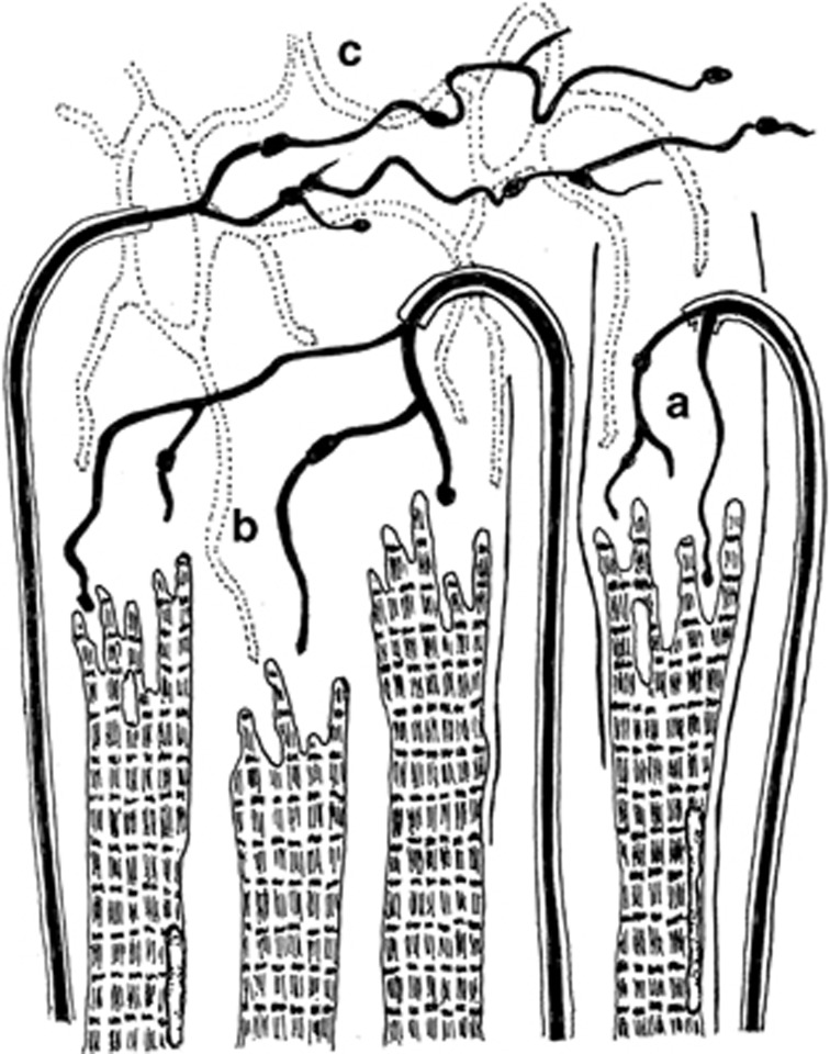
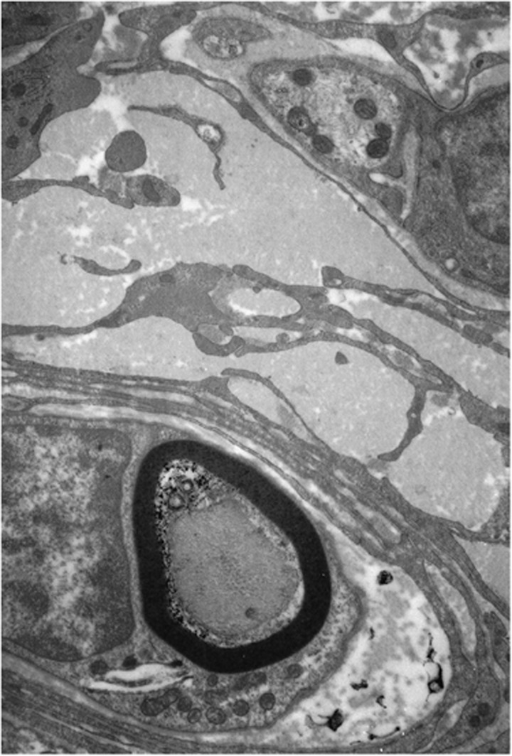
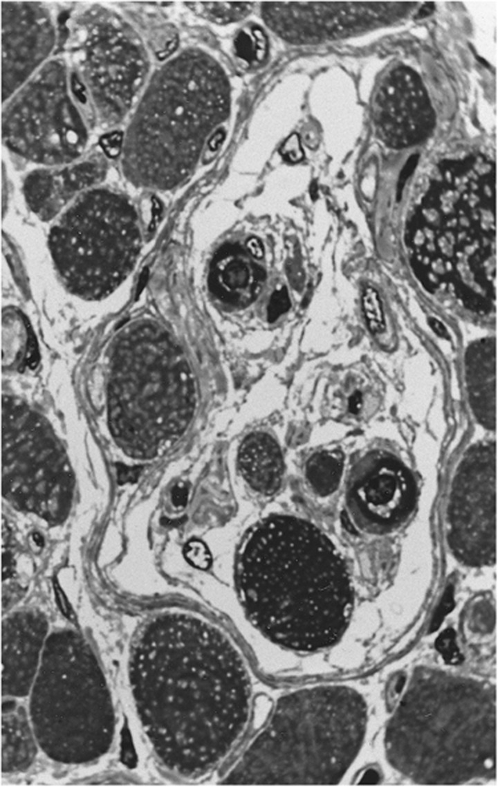
The sensory and motor control of human extraocular muscles (EOMs) have been subjected to considerable speculation in ophthalmic literature, often related to infranuclear structures such as the unique complement of muscle fibres and their associated sensory organs. The intrafusal fibres do not resemble their somatic counterparts and their peculiar morphology has raised questions about their proprioceptive capacity. No Golgi tendon organs have so far been observed and the myotendinous nerve endings, previously assumed to convey sensory information, have recently been argued to merely represent constituents of the efferent innervation serving the multiply innervated muscles fibres. These observations raise questions about the overall capacity to monitor the activity created by the generous efferent nerve supply observed in these muscles. Furthermore, the argued independent activity of muscular layers and compartments suggest that the required feedback must be highly structured and more specific than previously assumed. Yet, uncertainty about the source of such information remains. The purpose of this paper is to provide a short review of neuromuscular properties of human extraocular muscles. Their functional implications and the most reputable sources of proprioception will also be discussed. The promoted views are based on pertinent literature and previous research undertaken by the authors.
Figures
References
-
- Lienbacher K, Horn AK. Palisade endings and proprioception in extraocular muscles: a comparison with skeletal muscles. Biol Cybern. 2012;106:643–655. - PubMed
-
- Haugen IBK, Bruenech JR. The potential role of sensory receptors in ocular movements. Acta Ophthalmol Scand. 2007;85 (s240
-
- Ruskell GL. Extraocular muscle proprioceptors and proprioception. Prog Retin Eye Res. 1999;18 (3:269–291. - PubMed
-
- Demer JL, Oh SY, Poukens V. Evidence for active control of rectus extraocular muscle pulleys. Invest Ophthalmol Vis Sci. 2000;41 (6:1280–1290. - PubMed
Publication types
MeSH terms
LinkOut - more resources
Full Text Sources
Other Literature Sources
