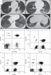
The lymphoid variant of hypereosinophilic syndrome: study of 21 patients with CD3-CD4+ aberrant T-cell phenotype
- PMID: 25398061
- PMCID: PMC4602413
- DOI: 10.1097/MD.0000000000000088
The lymphoid variant of hypereosinophilic syndrome: study of 21 patients with CD3-CD4+ aberrant T-cell phenotype
Abstract
The CD3-CD4+ aberrant T-cell phenotype is the most described in the lymphoid variant of hypereosinophilic syndrome (L-HES), a rare form of HES. Only a few cases have been reported, and data for these patients are scarce. To describe characteristics and outcome of CD3-CD4+ L-HES patients, we conducted a national multicentric retrospective study in the French Eosinophil Network. All patients who met the recent criteria of hypereosinophilia (HE) or HES and who had a persistent CD3-CD4+ T-cell subset on blood T-cell phenotyping were included. Clinical and laboratory data were retrospectively collected by chart review. CD3-CD4+ L-HES was diagnosed in 21 patients (13 females, median age 42 years [range, 5-75 yr]). Half (48%) had a history of atopic manifestations. Clinical manifestations were dermatologic (81%), superficial adenopathy (62%), rheumatologic (29%), gastrointestinal (24%), pulmonary (19%), neurologic (10%), and cardiovascular (5%). The median absolute CD3-CD4+ T-cell count was 0.35 G/L (range, 0.01-28.3), with a clonal TCRγδ rearrangement in 76% of patients. The mean follow-up duration after HES diagnosis was 6.9 ± 5.1 years. All patients treated with oral corticosteroids (CS) (n = 18) obtained remission, but 16 required CS-sparing treatments. One patient had a T-cell lymphoma 8 years after diagnosis, and 3 deaths occurred during follow-up.In conclusion, clinical manifestations related to CD3-CD4+ T cell-associated L-HES are not limited to skin, and can involve all tissue or organs affected in other types of HE. Contrary to FIP1L1-PDGFRA chronic eosinophilic leukemia patients, CS are always effective in these patients, but CS-sparing treatments are frequently needed. The occurrence of T-cell lymphoma, although rare in our cohort, remains a major concern during follow-up.
Conflict of interest statement
Financial support and conflicts of interest: This work was supported by 2 national Programme Hospitalier de Recherche Clinique, 2003 (RESEOPIL) and 2008 (HELPEO). The authors have no conflicts of interest to disclose.
Figures


References
-
- Bagot M, Bodemer C, Wechsler J, et al. [Non epidermotropic T lymphoma preceded for several years by hypereosinophilic syndrome]. Ann Dermatol Venereol. 1990;117:883–885. - PubMed
-
- Bank I, Amariglio N, Reshef A, et al. The hypereosinophilic syndrome associated with CD4+CD3- helper type 2 (Th2) lymphocytes. Leuk Lymphoma. 2001;42:123–133. - PubMed
-
- Baseggio L, Berger F, Morel D, et al. Identification of circulating CD10 positive T cells in angioimmunoblastic T-cell lymphoma. Leukemia. 2006;20:296–303. - PubMed
-
- Bergua JM, Prieto-Pliego E, Roman-Barbera A, et al. Resolution of left and right ventricular thrombosis secondary to hypereosinophilic syndrome (lymphoproliferative variant) with reduced intensity conditioning allogenic stem cell transplantation. Ann Hematol. 2008;87:937–938. - PubMed
-
- Brugnoni D, Airo P, Rossi G, et al. A case of hypereosinophilic syndrome is associated with the expansion of a CD3-CD4+ T-cell population able to secrete large amounts of interleukin-5. Blood. 1996;87:1416–1422. - PubMed
Publication types
MeSH terms
Substances
LinkOut - more resources
Full Text Sources
Other Literature Sources
Research Materials
Miscellaneous

