

T-DM1, a novel antibody-drug conjugate, is highly effective against uterine and ovarian carcinosarcomas overexpressing HER2
- PMID: 25398397
- PMCID: PMC4310789
- DOI: 10.1007/s10585-014-9688-8
T-DM1, a novel antibody-drug conjugate, is highly effective against uterine and ovarian carcinosarcomas overexpressing HER2
Abstract
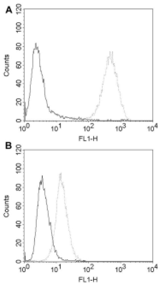
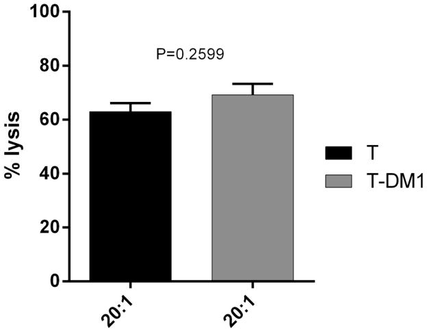
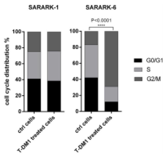
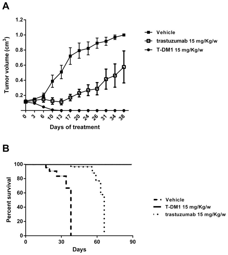
Ovarian and uterine carcinosarcoma (CS) are characterized by their aggressive clinical behavior and poor prognosis. We evaluated the efficacy of trastuzumab-emtansine (T-DM1), against primary HER2 positive and HER2 negative CS cell lines in vitro and in vivo. Eight primary CS cell lines were evaluated for HER2 amplification and protein expression by fluorescence in situ hybridization, immunohistochemistry, flow cytometry and qRT-PCR. Sensitivity to T-DM1-induced antibody-dependent-cell-mediated-cytotoxicity (ADCC) was evaluated in 4-h-chromium-release-assays. T-DM1 cytostatic and apoptotic activities were evaluated using flow cytometry based proliferation assays. In vivo activity of T-DM1 was also evaluated. HER2 protein overexpression and gene amplification were detected in 25 % (2/8) of the primary CS cell lines. T-DM1 and T were similarly effective in inducing strong ADCC against CS overexpressing HER2 at 3+ levels. In contrast, T-DM1 was dramatically more effective than T in inhibiting cell proliferation (P < 0.0001) and in inducing G2/M phase cell cycle arrest in the HER2 expressing cell lines (shift of G2/M: mean ± SEM from 14.87 ± 1.23 to 66.57 ± 4.56 %, P < 0.0001). Importantly, T-DM1 was highly active at reducing tumor formation in vivo in CS xenografts overexpressing HER2 (P = 0.0001 and P < 0.0001 compared to T and vehicle respectively) with a significantly longer survival when compared to T and vehicle mice (P = 0.008 and P = 0.0001 respectively). T-DM1 may represent a novel treatment option for the subset of HER2 positive CS patients with disease refractory to chemotherapy.
Conflict of interest statement
The authors declare that they have no competing interests.
Figures

References
-
- D’Angelo E, Prat J. Pathology of mixed Müllerian tumours. Hum Pathol Best Pract Res Clin Obstet Gynaecol. 2011;25:705–18. - PubMed
-
- Brooks SE, Zhan M, Cote T, Baquet CR. Surveillance, epidemiology, and end results analysis of 2677 cases of uterine sarcoma 1989–1999. Gynecol Oncol. 2004;93:204–8. - PubMed
-
- Garg G, Shah JP, Kumar S, Bryant CS, Munkarah A, Morris RT. Ovarian and uterine carcinosarcomas: a comparative analysis of prognostic variables and survival outcomes. Int J Gynecol Cancer. 2010;20:888–894. - PubMed
-
- Mano MS, Rosa DD, Azambuja E, Ismael G, Braga S, D’Hondt V, Piccart M, Awada A. Current management of ovarian carcinosarcoma. Int J Gynecol Cancer. 2007;17:316–24. - PubMed
Publication types
MeSH terms
Substances
Grants and funding
LinkOut - more resources
Full Text Sources
Other Literature Sources
Medical
Research Materials
Miscellaneous
