. 2015 Mar;29(3):786-95.
doi: 10.1096/fj.14-254284. Epub 2014 Nov 14.

The transcriptional corepressor MTGR1 regulates intestinal secretory lineage allocation

Affiliations

The transcriptional corepressor MTGR1 regulates intestinal secretory lineage allocation

Bobak Parang et al. FASEB J. 2015 Mar.

Abstract

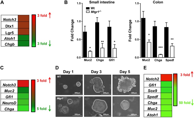
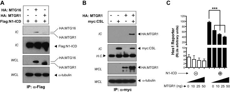
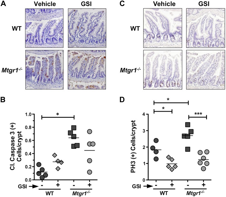
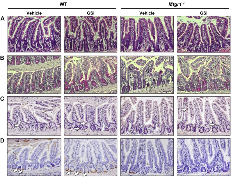
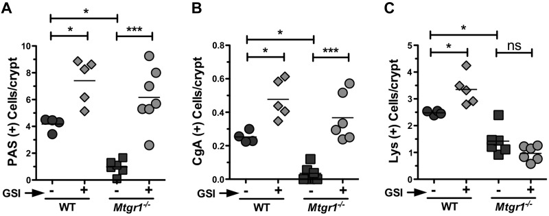
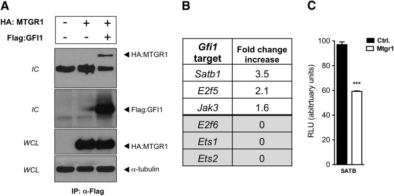
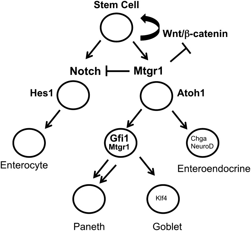
Notch signaling largely determines intestinal epithelial cell fate. High Notch activity drives progenitors toward absorptive enterocytes by repressing secretory differentiation programs, whereas low Notch permits secretory cell assignment. Myeloid translocation gene-related 1 (MTGR1) is a transcriptional corepressor in the myeloid translocation gene/Eight-Twenty-One family. Given that Mtgr1(-/-) mice have a dramatic reduction of intestinal epithelial secretory cells, we hypothesized that MTGR1 is a key repressor of Notch signaling. In support of this, transcriptome analysis of laser capture microdissected Mtgr1(-/-) intestinal crypts revealed Notch activation, and secretory markers Mucin2, Chromogranin A, and Growth factor-independent 1 (Gfi1) were down-regulated in Mtgr1(-/-) whole intestines and Mtgr1(-/-) enteroids. We demonstrate that MTGR1 is in a complex with Suppressor of Hairless Homolog, a key Notch effector, and represses Notch-induced Hairy/Enhancer of Split 1 activity. Moreover, pharmacologic Notch inhibition using a γ-secretase inhibitor (GSI) rescued the hyperproliferative baseline phenotype in the Mtgr1(-/-) intestine and increased production of goblet and enteroendocrine lineages in Mtgr1(-/-) mice. GSI increased Paneth cell production in wild-type mice but failed to do so in Mtgr1(-/-) mice. We determined that MTGR1 can interact with GFI1, a transcriptional corepressor required for Paneth cell differentiation, and repress GFI1 targets. Overall, the data suggest that MTGR1, a transcriptional corepressor well characterized in hematopoiesis, plays a critical role in intestinal lineage allocation.

Keywords: CBFA2T2; Notch signaling; Paneth cells.


Figures

Figure 1.
Figure 1.
Notch signaling is hyperactive, and secretory differentiation markers are down-regulated in the setting of Mtgr1 loss. A) Heatmap of dysregulated Notch-associated genes in microarray from laser-captured small intestinal crypts from WT and Mtgr1−/− mice (17). B) qPCR of the indicated lineage markers in small intestines (left) and colons (right) of WT (n = 4) and Mtgr1−/− (n = 4) mice. C) Heatmap of secretory-associated transcripts in WT and Mtgr1−/− MSIE cells. D) Representative images of enteroid cultures from WT and Mtgr1−/− mice 1, 3, and 5 d after plating. E) Heatmap of Notch-associated genes in WT and Mtgr1−/− small intestinal-derived enteroids. Red denotes increased fold expression relative to WT, and green denotes decreased fold expression relative to WT. The statistical test used was the Student’s t test. *P < 0.05; **P < 0.01; ***P < 0.001.
Figure 2.
Figure 2.
MTGR1 interacts with CSL and suppresses Notch-induced transcriptional activity. A) HA-epitope–tagged MTGR1 and Flag-epitope–tagged N1-ICD were transiently expressed in COS7L cells. HA:MTGR1 was blotted for in immune complexes (IC) (lane 3). HA-MTG16 and Flag-N1-ICD were also transiently transfected and immunoprecipitated as a positive control (lane 2). B) HA:MTGR1 and myc-epitope–tagged CSL were transiently expressed in COS7L cells in the combinations shown. The presence of myc:CSL and HA:MTGR1 was determined in immune complexes. C) MTGR1, in the quantities shown, was transiently expressed in NIH 3T3 cells with or without N1-ICD coexpression, along with the transcriptional reporter Hes1-Lux. Luciferase activity was quantified in whole-cell extracts by luminometry. Results were normalized to an internal Renilla constitutive reporter. The statistical test used was the Student’s t test. H.C., heavy chain; WCL, whole-cell lysate. ***P < 0.001.
Figure 3.
Figure 3.
GSI reduces proliferation but has no effect on apoptosis in the Mtgr1−/− intestine. Representative sections are shown of distal intestine (ileum) from vehicle or GSI-treated WT and Mtgr1−/− mice stained for (A) apoptosis marker cleaved caspase-3 or (C) proliferation marker pH3. Positive staining cells for (B) cleaved caspase-3 and (D) pH3 were quantified per crypt-villus unit. A minimum of 25 crypt-villus units was counted per mouse. The statistical test used was 1-way ANOVA. *P < 0.05; ***P < 0.001. Images were captured using a ×20 objective.
Figure 4.
Figure 4.
Notch inhibition increases goblet and enteroendocrine, but not Paneth, cells in Mtgr1−/− mice. Vehicle and GSI (DBZ, 10 μM/kg twice daily for 5 d)-treated WT and Mtgr1−/− mice are shown. A) H&E staining of distal intestine sections. PAS staining for goblet cells (B), CgA for enteroendocrine cells (C), and Lys staining for Paneth cells (D) in the distal intestine. White arrows indicate positive-staining cells. Images were captured using a ×20 objective.
Figure 5.
Figure 5.
Mtgr1 is required for Paneth lineage production after Notch inhibition. Quantification is shown of (A) PAS+, (B) CgA+, and (C) Lys+ cells per intestinal crypt/villus structure in WT and Mtgr1−/− mice treated with vehicle or GSI. A minimum of 25 crypt-villus units was counted per mouse. ns, not significant. The statistical test used was 1-way ANOVA. *P < 0.05; ***P < 0.001.
Figure 6.
Figure 6.
MTGR1 interacts with transcriptional corepressor GFI1 and can repress GFI1 targets. A) HA:MTGR1 and Flag:GFI1 were transiently expressed in COS7L cells. Flag:GFI1 was immunoprecipitated, and HA:MTGR1 was blotted for in immune complexes (IC). B) GFI1 repression targets Satb1, E2f5, and Jak3 were up-regulated in laser-captured crypts from WT and Mtgr1−/− mice (17). C) HA:MTGR1 was transfected into NIH 3T3 cells with the transcriptional reporter Satb1-Lux. Luciferase activity was quantified in whole-cell extracts by luminometry. Results were normalized to an internal Renilla constitutive reporter. Ctrl., control. The statistical test used was the Student’s t test. ***P < 0.001.
Figure 7.
Figure 7.
Schematic of MTGR1 regulating multiple branch points in intestinal lineage differentiation. We propose that MTGR1 acts at multiple levels in regulating intestinal epithelial differentiation. MTGR1 negatively regulates Wnt signaling by competing with β-catenin for TCF4 occupancy. As the stem cell divides and moves along the crypt-villus axis, MTGR1 acts as a negative regulator of Notch signaling by interacting with CSL and repressing Notch-induced transcriptional activity. As cells differentiate into either HES1+ enterocytes or ATOH1+ secretory progenitors, MTGR1 is also required downstream for Paneth cell differentiation potentially through its interaction with GFI1.

References

    1. Fre S., Huyghe M., Mourikis P., Robine S., Louvard D., Artavanis-Tsakonas S. (2005) Notch signals control the fate of immature progenitor cells in the intestine. Nature 435, 964–968 - PubMed
    1. Noah T. K., Donahue B., Shroyer N. F. (2011) Intestinal development and differentiation. Exp. Cell Res. 317, 2702–2710 - PMC - PubMed
    1. Bray S. J. (2006) Notch signalling: a simple pathway becomes complex. Nat. Rev. Mol. Cell Biol. 7, 678–689 - PubMed
    1. Kopan R., Ilagan M. X. (2009) The canonical Notch signaling pathway: unfolding the activation mechanism. Cell 137, 216–233 - PMC - PubMed
    1. Pellegrinet L., Rodilla V., Liu Z., Chen S., Koch U., Espinosa L., Kaestner K. H., Kopan R., Lewis J., Radtke F. (2011) Dll1- and dll4-mediated notch signaling are required for homeostasis of intestinal stem cells. Gastroenterology 140, 1230–1240, e1–e7 - PMC - PubMed

Publication types

MeSH terms

Substances