. 2014 Nov 15:14:9.
doi: 10.1186/s12899-014-0009-x.

Differential role of STIM1 and STIM2 during transient inward (T in) current generation and the maturation process in the Xenopus oocyte

Differential role of STIM1 and STIM2 during transient inward (T in) current generation and the maturation process in the Xenopus oocyte

Barbara Serrano-Flores et al. BMC Physiol. .

Abstract

Background: The Xenopus oocyte is a useful cell model to study Ca2+ homeostasis and cell cycle regulation, two highly interrelated processes. Here, we used antisense oligonucleotides to investigate the role in the oocyte of stromal interaction molecule (STIM) proteins that are fundamental elements of the store-operated calcium-entry (SOCE) phenomenon, as they are both sensors for Ca2+ concentration in the intracellular reservoirs as well as activators of the membrane channels that allow Ca2+ influx.

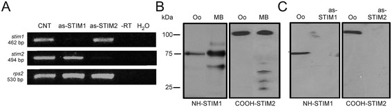
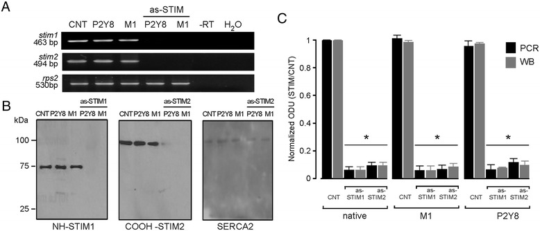
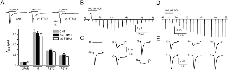
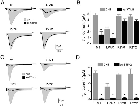
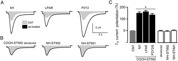
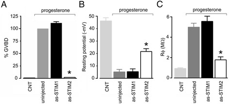
Results: Endogenous STIM1 and STIM2 expression was demonstrated, and their synthesis was knocked down 48-72 h after injecting oocytes with specific antisense sequences. Selective elimination of their mRNA and protein expression was confirmed by PCR and Western blot analysis, and we then evaluated the effect of their absence on two endogenous responses: the opening of SOC channels elicited by G protein-coupled receptor (GPCR)-activated Ca2+ release, and the process of maturation stimulated by progesterone. Activation of SOC channels was monitored electrically by measuring the T in response, a Ca2+-influx-dependent Cl- current, while maturation was assessed by germinal vesicle breakdown (GVBD) scoring and electrophysiology.

Conclusions: It was found that STIM2, but not STIM1, was essential in both responses, and T in currents and GVBD were strongly reduced or eliminated in cells devoid of STIM2; STIM1 knockdown had no effect on the maturation process, but it reduced the T in response by 15 to 70%. Thus, the endogenous SOCE response in Xenopus oocytes depended mainly on STIM2, and its expression was necessary for entry into meiosis induced by progesterone.


Figures

Figure 1
Figure 1
STIM expression in the Xenopus oocyte and its downregulation by as-STIM injection. A) shows the RT-PCR amplification of products that corresponded to the size expected for either stim1 or stim2 in native oocytes (CNT); the corresponding amplicons were absent in oocytes from the same batch that had been injected with either as-STIM1 or as-STIM2 48 h before the assay. The rps2 amplicon indicates the reaction efficiency, and -RT and H2O lanes correspond to negative controls, either RNA without RT, or to the reaction mix without a cDNA template, respectively. B) STIM1 and STIM2 were identified by Western blot analysis in protein extracts from oocytes (Oo) or mouse brain (MB, positive control) using either NH-STIM1 (left panel) or COOH-STIM2 (right panel) as antibody. C) A similar analysis as in B was made for batches of oocytes injected with H2O as control (CNT), or with as-STIM1 or as-STIM2 48 h before the protein extraction, in which cases proteins were eliminated. (in all cases 10 oocytes per condition).
Figure 2
Figure 2
Knockdown of STIM expression in oocytes co-injected with GPCR mRNA. A) RT-PCR amplification of stim1, stim2, or rps2 in batches of oocytes injected with H2O (CNT) or with cRNA (50 ng per oocyte) coding for either P2Y8 or M1 GPCR. In oocytes co-injected with as-STIM1 or as-STIM2 (50 ng per oocyte) together with P2Y8 or M1 cRNA, the corresponding STIM amplicon was downregulated. Control reactions illustrate specificity; rps2 amplicons are positive controls, and -RT and H2O lanes show negative controls. B) Similar groups of oocytes as in A) were assayed using the Western blot technique; in this case oocytes from the same donor injected with one GPCR mRNA (P2Y8 or M1) alone, or co-injected with as-STIM1, were tested with NH-STIM1, while as-STIM2-injected oocytes were probed with COOH-STIM2. In both as-STIM groups SERCA was used as gel-loading control. C) The graph shows the densitometric analysis of bands, summarizing the results obtained in different preparations of 10 oocytes per group and repeated in 3–5 frogs. Both PCR products and bands detected by Western blot (WB) were analyzed for batches of oocytes injected with H2O (CNT) or with either 50 ng as-STIM1 or as-STIM2 alone (native group). Similar analysis was made for batches of control oocytes injected with P2Y8 or M1 cRNA alone, and oocytes from the same frogs co-injected with either as-STIM or as-STIM together with the GPCR cRNA. Optical density units (ODU) for each band were normalized against the value obtained in the corresponding CNT conditions (*p < 0.01).
Figure 3
Figure 3
I osc and T in responses activated by agonist stimulation. A) Strength of I osc elicited by first agonist application did not change by knockdown of STIM1 or STIM2, compared with that obtained in CNT oocytes; top traces are typical responses elicited by ACh, similar responses were obtained by FBS or ATP applications, and the graph shows the average I osc responses obtained in oocytes held at −60 mV. B) Record illustrating the activation of T in current obtained in an oocyte expressing the M1 receptor by a single ACh (100 μM) application for 40 s (acute protocol). Oocytes were held at −10 mV while being superfused with NR solution and stepped to −100 mV for 4 s every 40 s; sudden hyperpolarization generated T in current responses that follow consistent kinetics with a peak amplitude response at 280–360 s (c); after that the response was washed out with a similar time course. C) Shows the T in current during the steps from −10 to −100 mV indicated with letters in panel B). D) A similar T in current response elicited in an oocyte from the same frog that was pre-incubated with 1 μM ACh for 4 h (long-lasting protocol), then monitored with the same electrical recording parameters and stimulated with 100 μM ACh. E) Shows the T in responses indicated with the same letters as in D). In this protocol T in current was consistently activated from the beginning of the record, and a transient inhibition of the response was noted during application of the agonist (b); after that, T in recovered and remained fully activated for a long period of time. Similar responses were obtained using oocytes expressing P2Y receptors and stimulating with ATP.
Figure 4
Figure 4
Specific STIM knockdown by oocyte injection of as-STIM differentially decreased the T in current. A) Oocytes induced to express M1, P2Y8, or P2Y2 receptors were stimulated with either ACh or ATP (100 μM), and LPAR in native oocytes were stimulated by FBS (1:1000 dilution); the resulting T in currents (CNT, gray areas) were compared with the T in obtained in oocytes from the corresponding group that were also injected with 50 ng as-STIM1 (superimposed black traces); all responses were monitored 48–72 h after oocyte injection. B) The graph shows the results obtained using the different experimental conditions illustrated in A). C) In a set of experiments similar to those shown in A), T in currents were monitored, and the peak amplitudes of non-injected CNT oocytes were compared with those of oocytes injected (48–72 h before recording) with 50 ng as-STIM2 and stimulated with the agonists. D) The graph shows the results obtained using the different experimental conditions illustrated in C). Bars correspond to the mean (± SEM) of the T in peak amplitude of 10–15 oocytes from 5–6 frogs (*p < 0.01, as-STIM vs. CNT).
Figure 5
Figure 5
Oocyte injection with COOH-STIM2 antibody produced a strong potentiation of T in current response. A) T in current responses were monitored in two conditions: non-loaded oocytes (CNT) and oocytes loaded with COOH-STIM2 antibody (ab-loaded). T in responses were elicited by ACh, FBS, or ATP application, depending on the receptor to be stimulated. In all cases, a strong potentiation of the response was observed in ab-loaded oocytes. B) Oocytes stimulated by ACh (M1) loaded with denatured COOH-STIM2 had control-like responses, while NH-STIM2 or NH-STIM1 loading did not produce T in potentiation. C) The graph shows the results obtained using the different experimental conditions illustrated in A and B; each bar corresponds to the mean (± SEM) of the T in peak amplitude normalized against the CNT current of 10–15 oocytes from 3–6 frogs (*p < 0.01).
Figure 6
Figure 6
Effect of as-STIM2 on GVBD and oocyte membrane characteristics during maturation induced by progesterone. A) The maturation process promoted by progesterone (10 μM) was analyzed in uninjected oocytes, or in oocytes injected 72 h prior to the assay with either as-STIM1 or as-STIM2, and compared with control oocytes in the absence of progesterone. GVBD was quantified after 8–12 h in presence of progesterone (10 oocytes per group, repeated using 3 different frogs) and is normalized against the value observed in uninjected oocytes. B) Resting membrane potential was monitored 8–12 h after addition of progesterone in the same groups of oocytes (n = 3-5, repeated in 3 frogs) as in A). C) The input membrane resistance (Rϕ) was estimated over the range from −80 to −20 mV in the different oocyte groups treated in the same conditions. Control groups, without progesterone, included both uninjected and antisense-injected oocytes. In all cases, values for as-STIM2-injected groups were different from as-STIM1-injected or uninjected groups (*p < 0.01).

Similar articles

Cited by

References

    1. Berridge MJ, Lipp P, Bootman MD. The versatility and universality of calcium signalling. Nat Rev Mol Cell Biol. 2000;1:11–21. doi: 10.1038/35036035. - DOI - PubMed
    1. Parker I, Miledi R. Changes in intracellular calcium and in membrane currents evoked by injection of inositol trisphosphate into Xenopus oocytes. Proc R Soc Lond B Biol Sci. 1986;228:307–315. doi: 10.1098/rspb.1986.0057. - DOI - PubMed
    1. Yao Y, Tsien R. Calcium current activated by depletion of calcium stores in Xenopus oocytes. J Gen Physiol. 1997;109:703–715. doi: 10.1085/jgp.109.6.703. - DOI - PMC - PubMed
    1. Hartzell HC: Activation of different Cl−currents in Xenopus oocytes by Ca2+liberated from stores and by capacitative Ca2+influx.J Gen Physiol 1996, 108:157–175. - PMC - PubMed
    1. Parekh AB, Penner R. Store depletion and calcium influx. Physiol Rev. 1997;77:901–930. - PubMed

Publication types

LinkOut - more resources