. 2014 Dec;35(12):1556-65.
doi: 10.1038/aps.2014.88. Epub 2014 Nov 17.

Probucol suppresses human glioma cell proliferation in vitro via ROS production and LKB1-AMPK activation

Affiliations

Probucol suppresses human glioma cell proliferation in vitro via ROS production and LKB1-AMPK activation

Yong-sheng Jiang et al. Acta Pharmacol Sin. 2014 Dec.

Retraction in

Abstract

Aim: Probucol, an anti-hyperlipidemic drug, has been reported to exert antitumor activities at various stages of tumor initiation, promotion and progression. In this study we examined whether the drug affected glioma cell growth in vitro and the underlying mechanisms.

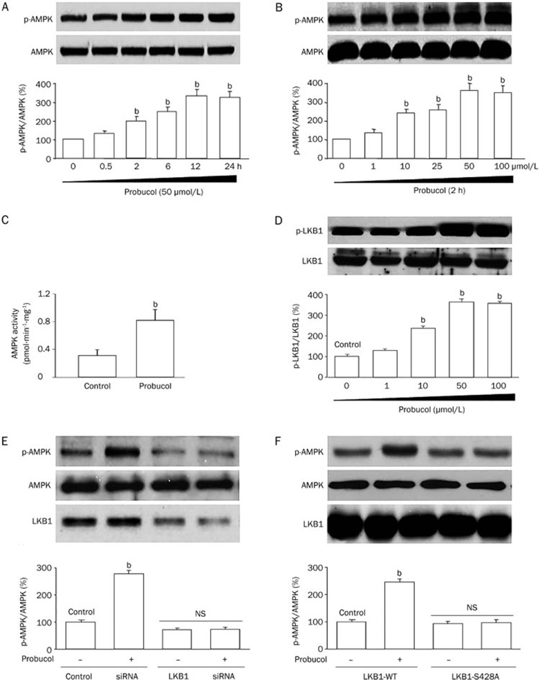
Methods: Human glioma U87 and glioblastoma SF295 cell lines were used. Cell proliferation was accessed using the cell proliferation assay and BrdU incorporation. The phosphorylation of AMPK, liver kinase B1 (LKB1) and p27(Kip1) was detected by Western blot. The activity of 26S proteasome was assessed with an in situ fluorescent substrate. siRNAs were used to suppress the expression of the relevant signaling proteins.

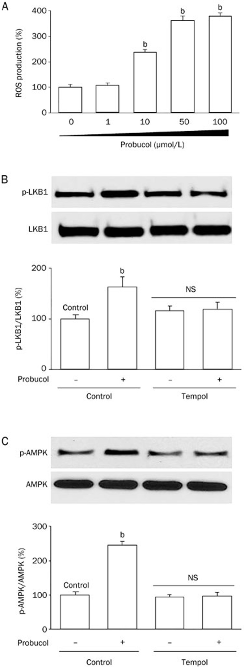
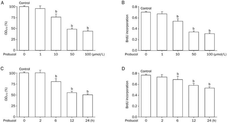
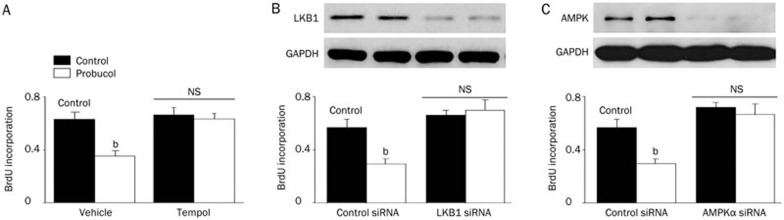
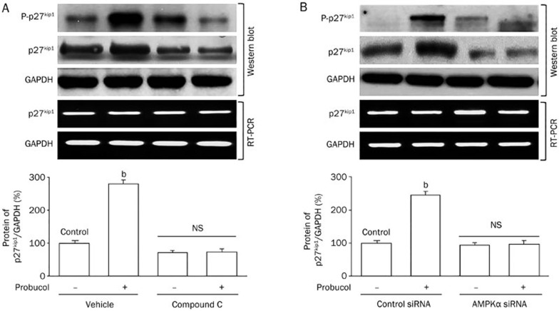
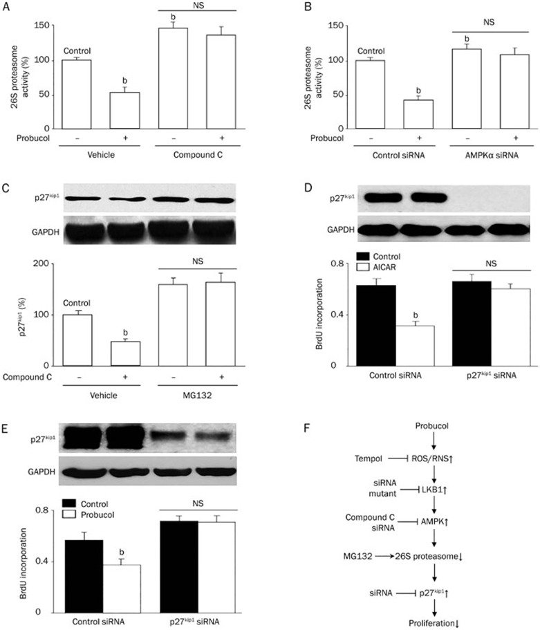
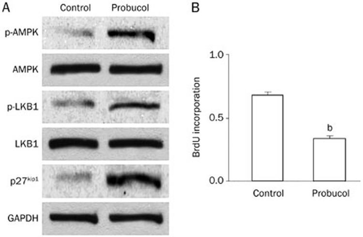
Results: Treatment of U87 glioma cells with probucol (10-100 μmol/L) suppressed the cell proliferation in dose- and time dependent manners. Meanwhile, probucol markedly increased the ROS production, phosphorylation of AMPK at Thr172 and LKB1 at Ser428 in the cells. Furthermore, probucol significantly decreased 26S proteasome activity and increased p27(Kip1) protein level in the cells in an AMPK-dependent manner. Probucol-induced suppression of U87 cell proliferation could be reversed by pretreatment with tempol (a superoxide dismutase mimetic), MG132 (proteasome inhibitor) or compound C (AMPK inhibitor), or by gene silencing of LKB1, AMPK or p27(Kip1). Similar results were observed in probucol-treated SF295 cells.

Conclusion: Probucol suppresses human glioma cell proliferation in vitro via ROS production and LKB1-AMPK activation, which reduces 26S proteasome-dependent degradation of p27(Kip1).


Figures

Figure 1
Figure 1
Probucol activates AMPK signaling via LKB1-mediated phosphorylation of Thr172 in cultured U87 glioma cells. (A) Time-dependent effects of probucol on AMPK-Thr172 phosphorylation in U87 glioma cells. Confluent U87 glioma cells were exposed to 50 μmol/L probucol as indicated. The blot is representative of three independent experiments. bP<0.05 vs control. (B) Dose-dependent effects of probucol on AMPK-Thr172 phosphorylation in U87 glioma cells. Confluent U87 glioma cells were exposed to probucol (1–100 μmol/L) for 2 h. The blot is representative of three independent experiments. bP<0.05 vs control. (C) Confluent U87 glioma cells were treated with vehicle or probucol (50 μmol/L) for 2 h. AMPK activity was assayed using the SAMS peptide as a substrate. n=5 in each group. bP<0.05 vs control. (D) Time-dependent effects of probucol on LKB1-Ser428 phosphorylation in U87 glioma cells. Confluent U87 glioma cells were exposed to 50 μmol/L probucol as indicated. The blot is representative of three independent experiments. bP<0.05 vs control. (E) U87 glioma cells were transfected with control siRNA or LKB1 siRNA for 48 h. The infected cells were then treated with probucol (50 μmol/L) for 2 h. (F) U87 glioma cells were infected with GFP-adenovirus or adenovirus expressing the mutant LKB1 (Ad-S428A) for 48 h. The infected cells were then treated with probucol (50 μmol/L) for 2 h. AMPK-Thr172 phosphorylation was detected by Western blot. The blot is representative from three independent experiments.
Figure 2
Figure 2
Reactive oxygen species (ROS) mediate probucol-induced AMPK activation in U87 glioma cells. (A) A confluent monolayer of U87 glioma cells were cultured in probucol (1–100 μmol/L) for 2 h. ROS production was assayed by DHE fluorescence. Data are expressed as the mean±SEM. n=5 in each group. bP<0.05 vs control. (B and C) Cultured U87 glioma cells were pre-incubated with tempol (1 mmol/L) for 30 min and then exposed to probucol (50 μmol/L) for 2 h. LKB1-Ser428 phosphorylation (B) and AMPK-Thr172 phosphorylation (C) were detected by Western blot. The blot is representative from three independent experiments. bP<0.05 vs control.
Figure 3
Figure 3
Probucol inhibits U87 glioma cell proliferation. (A) Cultured U87 glioma cells were incubated with probucol (1–100 μmol/L) for 2 h after an overnight serum starvation. The cell proliferation assay was performed following the manufacturer's protocol. (B) BrdU incorporation was measured in U87 glioma cells. (C) Cultured U87 glioma cells were incubated with 50 μmol/L probucol (2–24 h) after starvation overnight. Cell proliferation assay was performed as per the manufacturer's protocol. (D) BrdU incorporation was measured in U87 glioma cells. Mean±SEM. n=5. bP<0.05 vs control.
Figure 4
Figure 4
Probucol inhibits U87 glioma cell proliferation via LKB1/AMPK signaling. (A) Cultured U87 glioma cells pretreated with tempol (1 mmol/L) for 30 min were incubated with probucol (50 μmol/L) for 24 h. The cell proliferation assay was performed as the manufacturer's protocol. n=3 in each group. bP<0.05 vs control. NS indicates no significance. (B) Cultured U87 glioma cells transfected with LKB1 siRNA for 48 h were incubated with probucol (50 μmol/L) for 24 h. Cell proliferation assay was performed as the manufacturer's protocol. n=3 in each group. bP<0.05 vs control. NS indicates no significance. (C) Cultured U87 glioma cells transfected with AMPKα siRNA for 48 h were incubated with probucol (50 μmol/L) for 24 h. Cell proliferation assay was performed as the manufacturer's protocol. n=3. bP<0.05 vs control. NS indicates no significance.
Figure 5
Figure 5
Probucol increases p27Kip1 protein levels, but not gene expression, in glioma cells via AMPK activation. (A) Cultured U87 glioma cells pretreated with compound C (10 μmol/L) for 30 min were incubated with probucol (50 μmol/L) for 12 h. (B) Cultured U87 glioma cells transfected with AMPKα siRNA for 48 h were incubated with probucol (50 μmol/L) for 12 h. The phosphorylated and total p27Kip1 protein levels were assessed by Western blot analysis. The p27Kip1 mRNA level was assessed by RT-PCR analysis. Data are representative of 3 independent experiments. bP<0.05 vs control. NS indicates no significance.
Figure 6
Figure 6
Probucol suppress proliferation of glioma cells, mediated by the 26S proteasome and p27Kip1. (A) Cultured U87 glioma cells pretreated with compound C (10 μmol/L) for 30 min were incubated with probucol (50 μmol/L) for 12 h. The 26S proteasome activity was assessed by an in situ fluorescent substrate. bP<0.05 vs control. NS indicates no significance. (B) Cultured U87 glioma cells transfected with AMPKα siRNA for 48 h were incubated with probucol (50 μmol/L) for 12 h. The 26S proteasome activity was assessed by an in situ fluorescent substrate. bP<0.05 vs control. NS indicates no significance. (C) Cultured cells pretreated with MG132 (0.5 μmol/L) for 30 min were incubated with or without compound C (10 μmol/L) for 24 h. The total p27Kip1 protein level was assessed by Western blot analysis. This blot is representative blot from 3 independent experiments. bP<0.05 vs control. NS indicates no significance. (D and E) U87 glioma cells were transfected with either control or p27Kip1 siRNA for 48 h and then left untreated or treated with (D) AICAR (1 mmol/L) or (E) probucol (100 μmol/L) for 24 h. BrdU incorporation was used to measure U87 glioma cell proliferation. n=5 in each group. bP<0.05 vs control. NS indicates no significance. (F) Proposed molecular mechanism for the suppressive effects of probucol on U87 glioma cell proliferation.
Figure 7
Figure 7
Probucol activates LKB1-AMPK signaling, increases p27Kip1 protein levels, and reduces proliferation of human glioblastoma cells. SF295 cells were incubated with probucol (50 μmol/L) for 12 h. Western blotting of total cell lysates was performed to assay (A) protein levels of p-LKB1, p-AMPK, p27Kip1, and to determine (B) cell proliferation by BrdU incorporation. Mean±SEM. n=3 in each group. bP<0.05 vs control.

Similar articles

Cited by

References

    1. Ricard D, Idbaih A, Ducray F, Lahutte M, Hoang-Xuan K, Delattre JY. Primary brain tumours in adults. Lancet. 2012;379:1984–96. - PubMed
    1. Van Meir EG, Hadjipanayis CG, Norden AD, Shu HK, Wen PY, Olson JJ. Exciting new advances in neuro-oncology: the avenue to a cure for malignant glioma. CA Cancer J Clin. 2010;60:166–93. - PMC - PubMed
    1. Wong ML, Kaye AH, Hovens CM. Targeting malignant glioma survival signalling to improve clinical outcomes. J Clin Neurosci. 2007;14:301–8. - PubMed
    1. Jansen M, Ten Klooster JP, Offerhaus GJ, Clevers H. LKB1 and AMPK family signaling: the intimate link between cell polarity and energy metabolism. Physiol Rev. 2009;89:777–98. - PubMed
    1. Xie Z, Dong Y, Scholz R, Neumann D, Zou MH. Phosphorylation of LKB1 at serine 428 by protein kinase C-zeta is required for metformin-enhanced activation of the AMP-activated protein kinase in endothelial cells. Circulation. 2008;117:952–62. - PMC - PubMed

Publication types

MeSH terms

LinkOut - more resources