. 2014 Nov 17:5:5380.
doi: 10.1038/ncomms6380.

Separate neural representations for physical pain and social rejection

Affiliations

Separate neural representations for physical pain and social rejection

Choong-Wan Woo et al. Nat Commun. .

Abstract

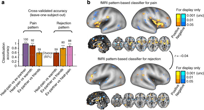
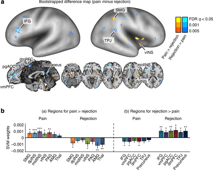
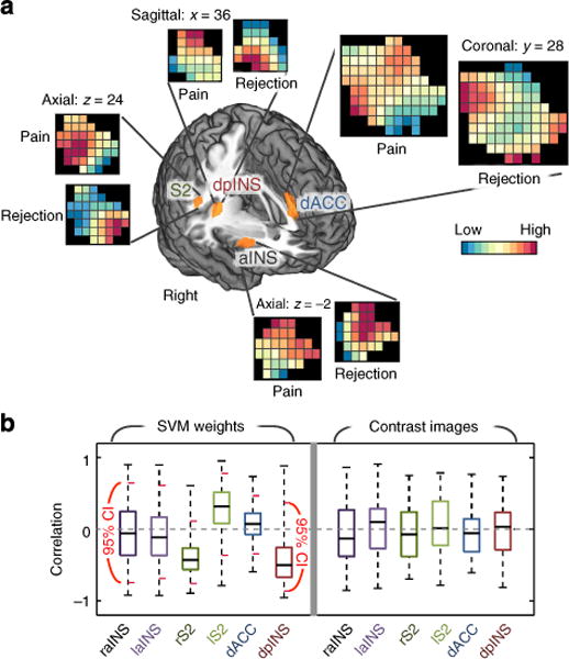
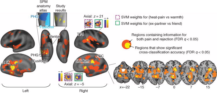
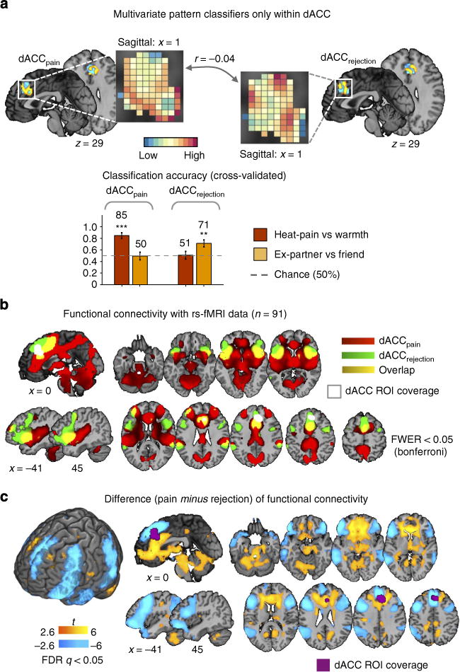
Current theories suggest that physical pain and social rejection share common neural mechanisms, largely by virtue of overlapping functional magnetic resonance imaging (fMRI) activity. Here we challenge this notion by identifying distinct multivariate fMRI patterns unique to pain and rejection. Sixty participants experience painful heat and warmth and view photos of ex-partners and friends on separate trials. FMRI pattern classifiers discriminate pain and rejection from their respective control conditions in out-of-sample individuals with 92% and 80% accuracy. The rejection classifier performs at chance on pain, and vice versa. Pain- and rejection-related representations are uncorrelated within regions thought to encode pain affect (for example, dorsal anterior cingulate) and show distinct functional connectivity with other regions in a separate resting-state data set (N = 91). These findings demonstrate that separate representations underlie pain and rejection despite common fMRI activity at the gross anatomical level. Rather than co-opting pain circuitry, rejection involves distinct affective representations in humans.


Conflict of interest statement

Competing financial interests: The authors declare no competing financial interests.

Figures

Figure 1
Figure 1. Behavioural results
(a) Experimental paradigm. The social rejection and somatic pain tasks each consisted of two consecutively administered runs of eight trials (that is, 16 total trials). The order of the two tasks was counterbalanced across participants. (i) Social rejection task: each trial in the social rejection task lasted 45 s and began with a 7-s fixation cross. Subsequently, participants saw a headshot photograph of their ex-partner (‘Ex-partner’ condition) or a close friend (‘Friend’ condition) for 15 s. A cue-phrase beneath each photo directed participants to think about how they felt during their break-up experience with their ex-partner or a specific positive experience with their friend. Subsequently, participants rated how they felt using a five-point scale. To reduce carryover effects between trials, participants then performed an 18-s visuo-spatial control task in which they saw an arrow pointing left or right and were asked to indicate which direction the arrow was pointing. Ex-partner versus Friend trials were randomly presented with the constraint that no trial repeated consecutively more than twice. (ii) Somatic pain task: the structure of somatic pain trials was identical to rejection trials with the following exceptions. During the 15-s thermal stimulation period, participants viewed a fixation cross and focused on the sensations they experienced during a hot (painful) or warm (non-painful) stimulus that was delivered (1.5-s temperature ramp up/down, 12 s at peak temperature) to their left volar forearm at temperatures calibrated for each person (for details, see Methods). They then rated the pain they experienced using a five-point scale. (b) Behavioural data from trial-by-trial pain and emotion rating (n = 60, eight trials for each condition). Error bars represent within-subject standard errors of the mean (s.e.m.). (c) Relative word frequency for negative emotion word categories among negative emotional words that participants used to describe the stream of thoughts while they were viewing the ex-partner’s photo in the scanner after the fMRI scanning. We used the Linguistic Inquiry and Word Count dictionary (LIWC) to categorize emotional words. ***P<0.001, multi-level generalized linear model.
Figure 2
Figure 2. Separate modifiability of fMRI pattern-based classifiers for pain and rejection
(a) Cross-validated (leave-one-subject-out) accuracy in two-choice classification tests (n = 59). The results demonstrated separate modifiability (each can be changed independent of the other) of the fMRI pattern-based classifiers. The dashed line indicates the chance level (50%), and the error bars represent standard error of the mean across subjects. (b) The distributed fMRI pattern maps in which voxel activity reliably contributes to the discrimination of pain (top panel) and rejection (bottom panel) from other conditions. The maps show thresholded voxel weights based on bootstrapping (10,000 samples) of SVMs for display only; all weights were used in classification. r between two pattern maps denotes Pearson’s correlation of voxel weights. ***P<0.001, binomial test.
Figure 3
Figure 3. The difference map between the fMRI pattern-based classifiers for pain and rejection
(a) The difference map in which values represent reliable differences between two discriminant weights (SVM weights for pain minus SVM weights for rejection) based on bootstrapping of SVMs (10,000 samples). The difference map is thresholded at q<0.05, FDR corrected. AMG, amygdala; dpINS, dorsal posterior insula; IFG, inferior frontal gyrus; midINS, middle insula; PAG, periaqueductal gray; pgACC, pregenual anterior cingulate cortex; preSMA, pre-supplementary motor area; SMG, supramarginal gyrus; Thal, thalamus; TPJ, temporal parietal junction; vINS, ventral insula; vmPFC, ventromedial prefrontal cortex. (b) Mean SVM weights for contiguous regions within the difference map (10,000 samples). The error bars represent standard error of the mean based on bootstrapping. *P<0.05, **P<0.01, ***P<0.001, one-sample t-test.
Figure 4
Figure 4. Multi-voxel pattern similarity analysis for pain-processing regions
(a) SVM classifier weight patterns within regions-of-interest (ROIs) from group-level GLM results. The regions were activated in both contrasts, Heat-pain versus Warmth and Ex-partner versus Friend, and have been implicated in both pain and rejection. Here, a liberal threshold (P<0.05, uncorrected) was applied to use large enough regions for multi-voxel pattern similarity analyses (the averaged number of voxels across six ROIs= 213). For GLM results corrected for multiple comparisons, see Supplementary Fig. 3. The patterns presented here are the averaged SVM classifier weights from bootstrap tests (10,000 samples). aINS, anterior insula; dACC, dorsal anterior cingulate cortex; dpINS, dorsal posterior insula; S2, secondary somatosensory cortex. (b) Left: the bootstrap test results for SVM classifier weight correlations. Right: the group-level correlations between fMRI activations of contrast images for pain and rejection (n = 59). The short red lines in the left panel indicate 95% confidence intervals obtained from bootstrap tests (10,000 samples). No regions showed significant correlations between SVM classifier weights, and no regions showed significant average correlations between patterns of contrast values across participants.
Figure 5
Figure 5. Cross-classification test results with local pattern classifiers
We conducted whole-brain searches for candidate regions for shared neural processes between pain and rejection with 6-mm radius spherical searchlights around centre voxels. To determine the candidate regions, we conducted cross-classification tests (with leave-one-subject-out cross-validation) among the regions that accurately classify both pain (Heat-pain versus Warmth) and rejection (Ex-partner versus Friend). The cross-classification test consisted of the following steps: (i) A local classifier was trained and cross-validated separately for one condition, and (ii) the classifier was tested on the other condition in the same leave-one-subject-out cross-validation test sample. Here we thresholded all results at FDR q<0.05 (P<0.0038) and visualized the 6-mm sphere coverage. PCC, posterior cingulate cortex; PHG, parahippocampal gyrus; RSC, retrosplenial cortex; TPJ, temporal parietal junction. The anatomical atlas was from the SPM anatomy toolbox.
Figure 6
Figure 6. Difference in functional connectivity patterns with dACC pattern classifiers for pain and rejection
(a) The multivariate pattern classifiers for pain and rejection within the dorsal anterior cingulate cortex (dACC) region-of-interest (ROI) (a top). The dACC ROI was defined by the searchlight analysis results presented in Fig. 5, which showed that the dACC ROI contained information for both pain and rejection conditions, but the patterns were distinct and non-transferrable. Using the dACC ROI, we trained linear SVMs to discriminate pain and rejection from their respective control conditions, and tested on out-of-sample participants. The pattern weights were uncorrelated with each other, r= −0.04. The cross-validated (leave-one-subject-out) accuracy in two-choice classification tests demonstrated separate modifiability of the pattern classifiers (a bottom). The dotted line indicates the chance level (= 50%), and the error bars represent standard error of the mean across subjects. **P<0.01, ***P<0.001, binomial test. (b) Seed-based functional connectivity with dACC pattern classifiers for pain and rejection. Here seeds were pattern expression values (the dot-product of a vectorized activation map and SVM weights within dACC) for pain and rejection. The functional connectivity for each condition was calculated with independent resting-state fMRI data (n = 91). These maps were thresholded at family-wise error rate (FWER)<0.05 using Bonferroni correction. Here we used Bonferroni correction instead of false discovery rate (FDR) because the latter provided too liberal thresholds for these functional connectivity patterns (uncorrected P<0.03) and therefore yielded non-sensible maps. (c) Paired t-test results between two seed-based functional connectivity patterns. The results were thresholded at FDR<0.05.

Comment in

References

    1. MacDonald G, Leary MR. Why does social exclusion hurt? The relationship between social and physical pain. Psychol Bull. 2005;131:202–223. - PubMed
    1. Eisenberger NI, Lieberman MD, Williams KD. Does rejection hurt? An fMRI study of social exclusion. Science. 2003;302:290–292. - PubMed
    1. Eisenberger NI. The pain of social disconnection: examining the shared neural underpinnings of physical and social pain. Nat Rev Neurosci. 2012;13:421–434. - PubMed
    1. Kross E, Berman MG, Mischel W, Smith EE, Wager TD. Social rejection shares somatosensory representations with physical pain. Proc Natl Acad Sci USA. 2011;108:6270–6275. - PMC - PubMed
    1. Novembre G, Zanon M, Silani G. Empathy for social exclusion involves the sensory-discriminative component of pain: a within-subject fMRI study. Soc Cogn Affect Neurosci. 2014 doi: 10.1093/scan/nsu038. - DOI - PMC - PubMed

Publication types