. 2015 Apr 15;523(6):869-91.
doi: 10.1002/cne.23711. Epub 2014 Dec 30.

Brain composition in Godyris zavaleta, a diurnal butterfly, Reflects an increased reliance on olfactory information

Affiliations

Brain composition in Godyris zavaleta, a diurnal butterfly, Reflects an increased reliance on olfactory information

Stephen H Montgomery et al. J Comp Neurol. .

Abstract

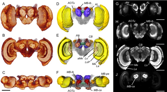
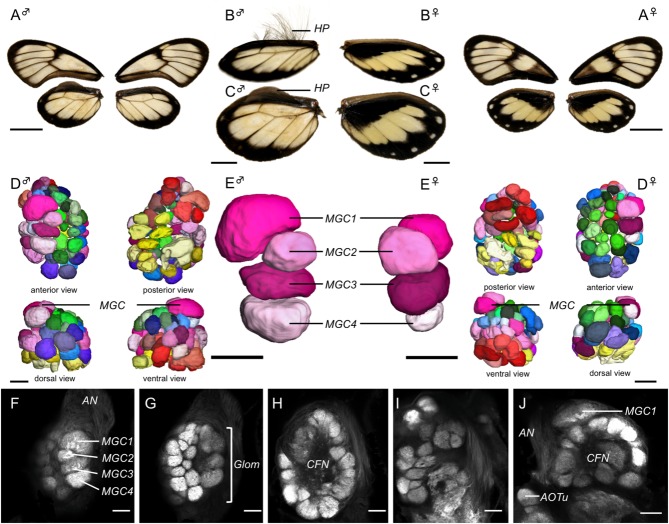
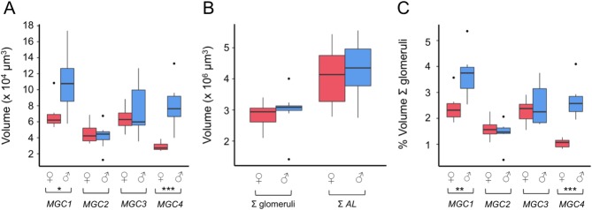
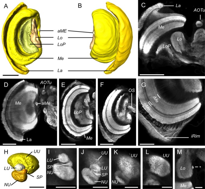
Interspecific comparisons of brain structure can inform our functional understanding of brain regions, identify adaptations to species-specific ecologies, and explore what constrains adaptive changes in brain structure, and coevolution between functionally related structures. The value of such comparisons is enhanced when the species considered have known ecological differences. The Lepidoptera have long been a favored model in evolutionary biology, but to date descriptions of brain anatomy have largely focused on a few commonly used neurobiological model species. We describe the brain of Godyris zavaleta (Ithomiinae), a member of a subfamily of Neotropical butterflies with enhanced reliance on olfactory information. We demonstrate for the first time the presence of sexually dimorphic glomeruli within a distinct macroglomerular complex (MGC) in the antennal lobe of a diurnal butterfly. This presents a striking convergence with the well-known moth MGC, prompting a discussion of the potential mechanisms behind the independent evolution of specialized glomeruli. Interspecific analyses across four Lepidoptera further show that the relative size of sensory neuropils closely mirror interspecific variation in sensory ecology, with G. zavaleta displaying levels of sensory investment intermediate between the diurnal monarch butterfly (Danaus plexippus), which invests heavily in visual neuropil, and night-flying moths, which invest more in olfactory neuropil. We identify several traits that distinguish butterflies from moths, and several that distinguish D. plexippus and G. zavaleta. Our results illustrate that ecological selection pressures mold the structure of invertebrate brains, and exemplify how comparative analyses across ecologically divergent species can illuminate the functional significance of variation in brain structure.

Keywords: Godyris zavaleta; Ithomiinae; Lepidoptera; adaptive brain evolution; antennal lobe; comparative neuroanatomy; macroglomerular complex; sexual dimorphism.


Figures

Figure 1
Figure 1
Overview of the anatomy of the G. zavaleta brain. A–C: Volume rendering of synapsin (3C11) immunofluorescence showing the surface morphology of the brain neuropil from the anterior (A), posterior (B), and dorsal (C) view. D–F: Surface reconstructions of the major neuropil compartments from the anterior (D), posterior (E), and dorsal (F) view. The midbrain houses the antennal lobes (AL) which lie at the base of the antennal nerve (AN in G), the anterior optic tubercles (AOTu), the central body (CB), protocerebral bridge (PB), and the mushroom body, which comprises the calyx (MB-ca), peduncle (MB-pe), and lobes (MB-lb); the optic lobes comprise the lamina (La), medulla (Me), and accessory medulla (aMe), the lobula (Lo), and lobula plate (LoP). G–J: Anti-synapsin immunofluorescence in frontal confocal sections taken at progressively more posterior levels. G: The antennal lobes (AL) are the most anterior midbrain neuropils. H: Further back, the anterior optic tubercles (AOTu) flank the mushroom body lobes (MB-lb), which occupy a dorsomedial position. I: The central body (CB) and the three main neuropils in the optic lobe, the lamina (La), medulla (Me), and lobula (Lo). J: The mushroom body calyx (MB-ca) at the back end of the midbrain. The individual displayed is female. Scale bars = 500 μm.
Figure 2
Figure 2
Sexual dimorphism in the wings and antennal lobes (AL). A–C: Wing morphology in males (♂) and females (♀) showing wing pigmentation on the dorsal surface (A) and the presence of hair-pencils (HP) on the hind wings of males from an oblique anterodorsal view (B) and a direct dorsal view (C). D,E: Surface reconstructions of the full complement of AL glomeruli (D), and of the subset of four glomeruli that form the macroglomerular complex (MGC1–4 in E). Among the ordinary glomeruli, green tones indicate the dorsalmost glomeruli, blue tones the middle layer, pink tones the posterior layer, red tones the posterodorsal glomeruli and yellow tones the posteromedial glomeruli. F–J: Synapsin immunofluorescence in single confocal sections of the AL. F–I: Frontal sections that move progressively deeper into the AL until reaching its posterior boundary. The four distinct glomeruli of the MGC (MGC1–4 in F) occupy the most anterior position in the AL, close to the root of the antennal nerve (AN in F). The ordinary glomeruli (Glom in G) surround the central fibrous neuropil (CFN in H,J). J: A horizontal section at the level of the anterior optic tubercle (AOTu) and shows MGC1 protruding from the general spherical shape of the AL. Scale bars = 1.0 cm in A; 0.5 cm in B,C; 50 μm in D–J.
Figure 3
Figure 3
Volumetric quantification of the sexual dimorphism in the antennal lobes. A: Raw volumes of MGC1–4 for males (♂) and females (♀). B: Raw volumes of the total volume of all glomeruli and the AL (including the CFN) for males (♂) and females (♀). C: Volumes of MGC1–4 for males (♂) and females (♀) as a proportion of the total volume of the glomeruli. The boxplots show the median (horizontal line in box), the interquartile range (range of the box), and the maximum and minimum of the range (whiskers); outliers are shown as separate data points. Significant differences between males and females are indicated by *P < 0.05, **P < 0.01, ***P < 0.001.
Figure 4
Figure 4
Anatomy of the visual neuropils. A,B: Surface reconstructions of the optic lobe neuropils viewed from posterior (A) and anterior (B). They comprise the lamina (La), the medulla (Me), and accessory medulla (aMe), the lobula (Lo), and the lobula plate (LoP). C–G: Synapsin immunofluorescence in single confocal sections of the optic lobe. C: A horizontal section showing all four major optic lobe neuropils (La, Me, Lo, LoP) together with the anterior optic tubercle (AOTu) in the midbrain. D–F: Frontal sections at increasing depths from anterior to posterior, beginning at a plane tangential through the lamina (La in D) and reaching the optic stalk (OS in F). G: The inner rim (iRim) of the lamina is a thin layer on its inner surface that is defined by intense synapsin immunofluorescence; it is also visible in C,D,F. Synapsin immunostaining also reveals the laminated structure of the medulla with two main subdivisions, the outer and inner medulla (oMe, iMe). H: Surface reconstruction of the AOTu from an oblique anterior view, showing the four component neuropils: the upper unit (UU), lower unit (LU), strap (SP), and the nodular unit (NU). I–L: Synapsin immunofluorescence in the AOTu in frontal confocal sections at increasing depths from anterior (I) to posterior (L). M: A small neuropil (asterisk) positioned at the medial margin of the lobula which may be homologous to the D. plexippus optic glomerular complex. Scale bars = 200 μm in A–F; 100 μm in G; 50 μm in H–M.
Figure 5
Figure 5
Anatomy of the mushroom body. A,B: Surface reconstruction of the mushroom body from the dorsal (A) and lateral (B) view. C–F: Synapsin immunofluorescence in horizontal confocal sections through the midbrain at increasing depths from dorsal towards ventral. The very dorsal (tangential) plane C shows the tips of the medial lobe (MB-lb) and of the y-lobe (y-lb). D: The main components of the lobes: the α-lb, y-lb, and the compartmentalized medial lobe (MB-lb). E: The Y-tract runs from the calyx (MB-ca) anteriorly to the lobes (MB-lb). F: Further ventral and level with the upper and lower units of the central body (CBU, CBL), the peduncles (Pe) provide the main connection between MB-ca and MB-lb. G–K: Details of mushroom body architecture as revealed by synapsin immunofluorescence in confocal sections. G: Frontal section showing the separate profiles of the Y-tract and pedunculus (MB-pe) in the central mid-brain; dorsal is up. H,I: Frontal sections through the calyx (Mb-ca). H: Each MB-ca consists of two fused calycal neuropils, which lack any obvious internal zonation. I: Emergence of the Y-tract at the anterodorsal boundary of MB-ca. J,K: Morphology of the partially merged dorsal lobe of the MB. Scale bars = 100 μm in A–F; 50 μm in G–K.
Figure 6
Figure 6
Anatomy of the central complex. A,B: Surface reconstruction of the central body from an anterior (A) and oblique anteroventral (B) view, showing the upper and lower subunit of the central body (CBU, CBL) and the three compartments of the noduli (No). C,D: Synapsin immunofluorescence in horizontal confocal sections showing the structure of the upper and lower CB (C) and the foliated dorsal surface of the lower CB (D). E: Surface reconstruction of the protocerebral bridge (PB) and posterior optic tubercles (POTu) from an oblique posterior view. F,G: Synapsin-immunofluorescence in frontal confocal sections showing the PB (F), and the POTu ventral to the MB-ca (G). H: The noduli (No) in a horizontal confocal section ventral to the CB showing three discrete compartments homologous to compartments II–IV in D. plexippus (Heinze and Reppert, 2012); the intensely stained compartment I, which partially encircles subcompartment II in D. plexippus was not clearly visible. Scale bars = 50 μm in A; 100 μm in C–F; 50 μm in G–H.
Figure 7
Figure 7
Principal component (PC) biplots of the relative size of major brain structures in four Lepidoptera. A: PC analysis of all neuropil. B: PC analysis excluding the optic lobe neuropil. In both cases the PC analysis was performed using residuals from a phylogenetically controlled regression between each neuropil and the rest of the total neuropil/total midbrain. Analysis using volumes expressed as percentages results in similar conclusions (Table4). The biplots show each species as a data point labeled with the first letter of the genus name (D = Danaus, G = Godyris, H = Heliothis, M = Manduca); vectors lengths are proportional to the variance of that variable/neuropil.

References

    1. Ali FA. Structure and metamorphosis of the brain and suboesophageal ganglion of Pieris brassicae (L.) (Lepidoptera: Pieridae) Trans R Entomol Soc Lond. 1974;125:363–412.
    1. Andersson S, Dobson HEM. Behavioral foraging responses by the butterfly Heliconius melpomene to Lantana camara floral scent. J Chem Ecol. 2003;29:2303–2318. - PubMed
    1. Andersson J, Borg-Karlson A-K, Vongvanich N, Wiklund C. Male sex pheromone release and female mate choice in a butterfly. J Exp Biol. 2007;210:964–970. - PubMed
    1. Barton RA, Harvey PH. Mosaic evolution of brain structure in mammals. Nature. 2000;405:1055–1058. - PubMed
    1. Barton RA, Purvis A, Harvey PH. Evolutionary radiation of visual and olfactory brain systems in primates, bats and insectivores. Philos Trans R Soc Lond B Biol Sci. 1995;348:381–392. - PubMed

Publication types

LinkOut - more resources