. 2014 Nov 17:5:5496.
doi: 10.1038/ncomms6496.

BRCA1 haploinsufficiency for replication stress suppression in primary cells

Affiliations

BRCA1 haploinsufficiency for replication stress suppression in primary cells

Shailja Pathania et al. Nat Commun. .

Abstract

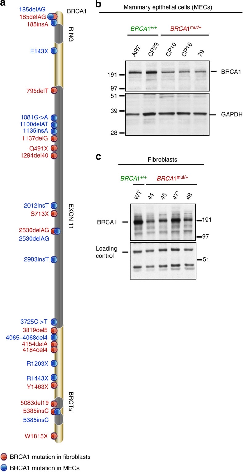
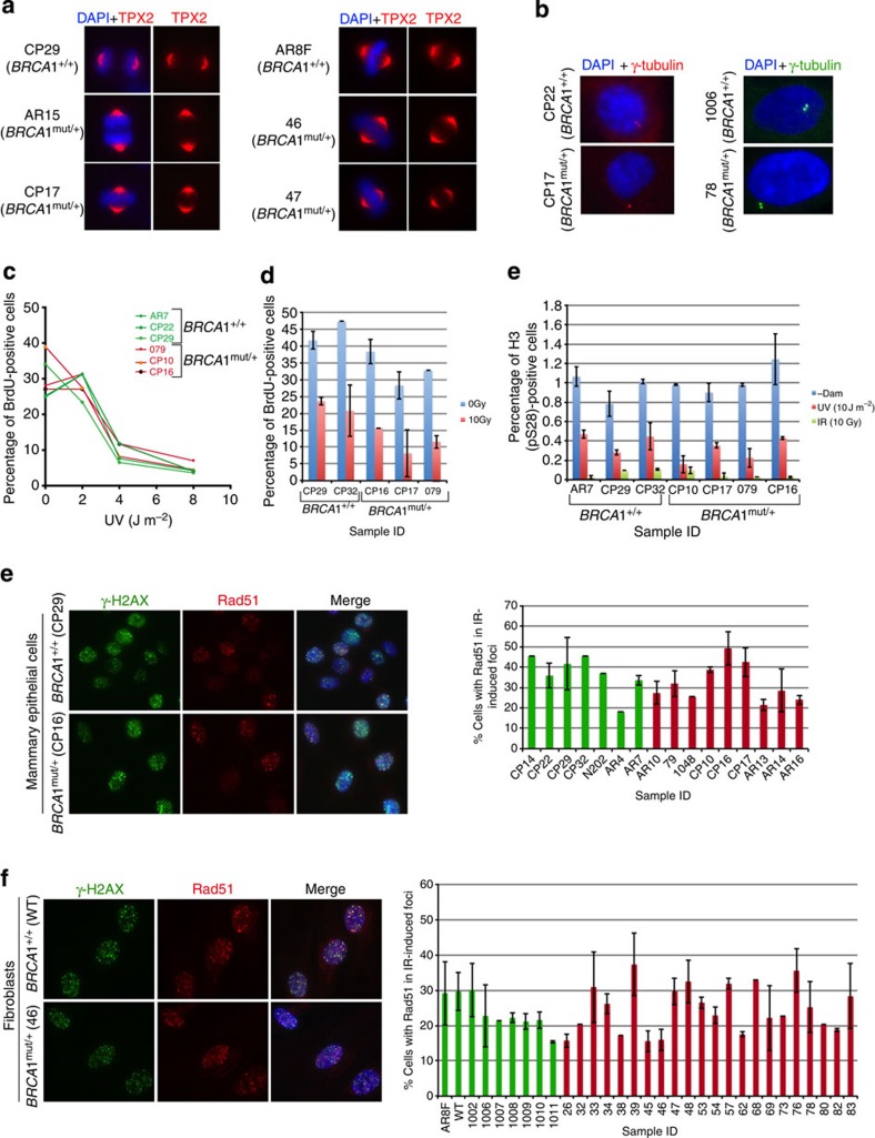
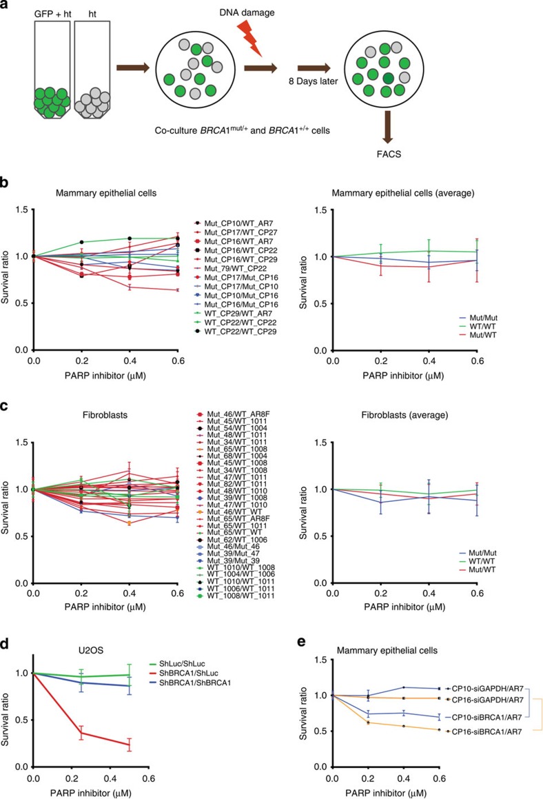
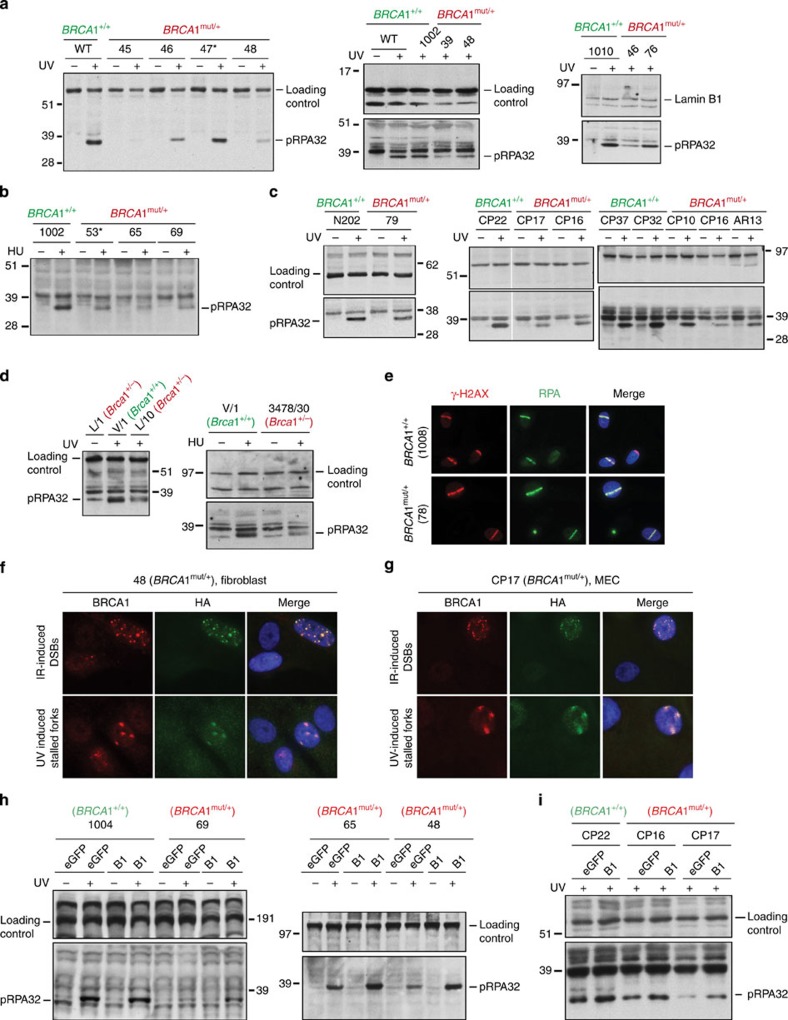
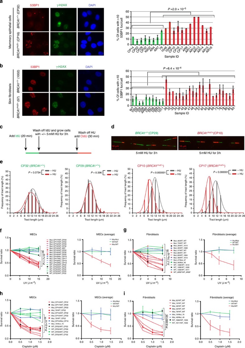
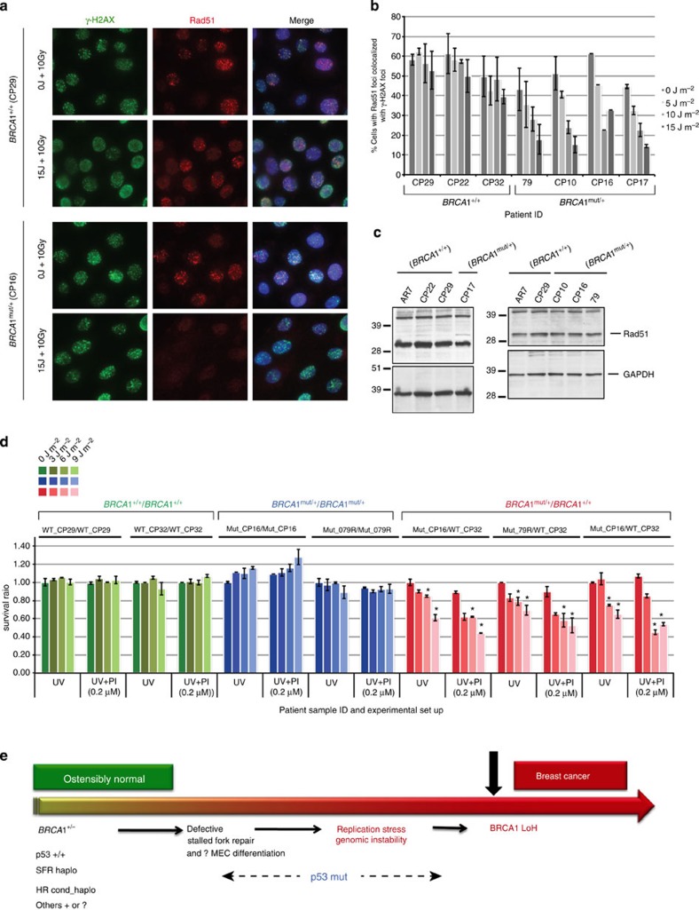
BRCA1-a breast and ovarian cancer suppressor gene-promotes genome integrity. To study the functionality of BRCA1 in the heterozygous state, we established a collection of primary human BRCA1(+/+) and BRCA1(mut/+) mammary epithelial cells and fibroblasts. Here we report that all BRCA1(mut/+) cells exhibited multiple normal BRCA1 functions, including the support of homologous recombination- type double-strand break repair (HR-DSBR), checkpoint functions, centrosome number control, spindle pole formation, Slug expression and satellite RNA suppression. In contrast, the same cells were defective in stalled replication fork repair and/or suppression of fork collapse, that is, replication stress. These defects were rescued by reconstituting BRCA1(mut/+) cells with wt BRCA1. In addition, we observed 'conditional' haploinsufficiency for HR-DSBR in BRCA1(mut/+) cells in the face of replication stress. Given the importance of replication stress in epithelial cancer development and of an HR defect in breast cancer pathogenesis, both defects are candidate contributors to tumorigenesis in BRCA1-deficient mammary tissue.


Figures

Figure 1
Figure 1. Distribution of BRCA1 mutations and BRCA1 protein in cells derived from BRCA1 mutation carriers.
(a) Cells were derived from skin punch biopsies and prophylactic mastectomies performed on BRCA1 mutation carrying women. (b) Western blot analysis of total BRCA1 protein levels in BRCA1mut/+ and BRCA1+/+ HMEC lines. Equivalent amounts of whole-cell lysate (prepared in NETN300) were electrophoresed, blotted and the blots probed with an anti-BRCA1 monoclonal Ab (SD118). GAPDH served as a loading control. (c) Western blot analysis of BRCA1 protein levels in the nuclear fraction of BRCA1mut/+ and BRCA1+/+ fibroblast strains. Cells were pre-lysed in pre-extraction buffer (PEB, details in Materials and Methods), and the pellet was re-suspended in NETN400 buffer to prepare a nuclear extract. The intense BRCA1 band in 47 (185delAG, marked by an asterisk) is likely the previously discovered truncated product of this mutant allele. A non- specific band served as the loading control.
Figure 2
Figure 2. Spindle pole formation, centrosome number, checkpoint activation and Rad51 recruitment to DSB.
(a) Representative images of HMECs (left panel) and skin fibroblasts (right panel), from BRCA1mut/+ and BRCA1+/+ were immunostained with an anti- TPX2 Ab to detect spindles; n=50 spindles were analysed for each strain. A summary of all strains that were tested in this assay is listed in supplementary Table 1. (b) Centrosome number was determined by immunostaining HMECs (left panel) and fibroblasts (right panel) with Ab to γ-tubulin; n=50 cells for each line were counted and cells with centrosomes ≤2 were considered normal. A summary of the lines that were tested is presented in Supplementary Table 1. (c) S-phase checkpoint in response to UV- and IR-induced DNA damage in control and BRCA1mut/+ strains. Three BRCA1+/+ (AR7, CP22 and CP29) and three BRCA1mut/+ HMEC strains (79, CP10 and CP16) were irradiated with increasing doses of UV (left panel). For IR-induced S-phase checkpoint analysis (right panel), cells were exposed to IR (10 Gy, red). Non-irradiated cells (0 Gy, blue) served as controls. Error bars indicate the s.d. between the results of three, independent experiments. (d) G2/M checkpoint activation in response to UV- and IR- induced DNA damage in BRCA1mut/+ and control cells. BRCA1+/+ and BRCA1mut/+ cells were irradiated with either UV (10 J m−2) or IR (10 Gy), allowed to recover for 2 h and then harvested for FACS analysis. The percentage of cells in mitosis was determined by staining cells with propidium iodine (PI) and phosphorylated histone H3 (S28) antibody. Mock-irradiated (-Dam) cells served as controls. (e) HMECs and (f) fibroblasts were exposed to IR (10 Gy) and allowed to recover for 4 h. Cells were fixed and co-immunostained with Abs to γ-H2AX and Rad51. Graphs depicting the fraction of cells with Rad51 foci that co-localized with γ-H2AX foci for each line are plotted for both HMECs and fibroblasts (right panels in e and f). Mean and s.d. of at least three experiments for each strain are shown. wt BRCA1+/+ (green) and BRCA1mut/+ (red) lines.
Figure 3
Figure 3. FACS-based cell survival assay shows that HR-DSBR is not defective in BRCA1mut/+ cells.
(a) FACS-based cell survival assay was used to determine the sensitivity of cells to various DNA damage inducing agents. BRCA1mut/+ and BRCA1+/+ ‘colour-coded’ cells were co-plated and exposed to DNA damaging agents. Cell survival data are plotted as a ratio of GFP positive to GFP negative cells. Ratio between WT/WT(Green), Mutant/Mutant (Blue) and Mutant/WT (Red) is plotted in the graphs below. (b) Combinations of BRCA1mut/+ and BRCA1+/+ HMECs were exposed to different concentrations of a PARP inhibitor, and the ratio of each of these combinations was plotted (left). The average ratio of WT/WT, Mut/Mut and Mut/WT was also calculated and plotted (right). (c) (Left) Combinations of BRCA1mut/+ and BRCA1+/+ fibroblasts were exposed to different concentrations of a PARP inhibitor, and the survival ratio of each of these combinations was plotted (left). An average ratio of WT/WT, Mut/Mut and Mut/WT was also calculated and plotted (right). (d) U20S cells (containing or lacking a GFP reporter) were infected with ShLuc (control) or ShBRCA1 coding lentiviral vectors. Green=ratio of number of ShLuc-treated cells to ShLuc-treated cells, that is (ShLuc/ShLuc), Blue=ratio of number of ShBRCA1-treated cells to ShBRCA1 treated-cells, that is, ShBRCA1/ShBRCA1 and Red=ratio of number of ShBRCA1-treated cells to ShLuc-treated cells, that is, ShBRCA1/ShLuc. Averages of the results of individual experiments are plotted. (e) BRCA1mut/+ (CP10 and CP16) were transduced with shRNA directed at GAPDH (siGAPDH) or BRCA1 (siBRCA1). Three days post transfection, combinations of siGAPDH or siBRCA1-transduced BRCA1mut/+ HMECs (CP10 and CP16) were co-plated with AR7 (a BRCA1+/+ HMEC) and exposed to various doses of a PARP inhibitor. Averages of the results generated by these combinations were plotted. Error bars were calculated as the standard error propagation (SEP) in the ratios of each of the combinations in three independent experiments.
Figure 4
Figure 4. BRCA1mut/+ cells derived from human and mouse tissue are defective in the generation of phospho-RPA32-coated ssDNA.
(a) Phospho-RPA32 (pRPA32) loading on chromatin is BRCA1 dependent. After UV-induced DNA damage, BRCA1mut/+ fibroblasts exhibited reduced pRPA32 loading on ssDNA, compared with BRCA1+/+ lines. Cells were irradiated with 30 J m−2 of UV and harvested 3 h post damage. Chromatin extracts were prepared, and the relevant western blot was probed with an antibody to phosphorylated RPA32 (S4/S8). The replication status for each line was tested on the day of the experiment by BrdU uptake measurement, and only those lines that exhibited similar replication profiles were analysed. A subset of lines tested is shown here. Western blots for other WT and BRCA1 mutant lines are shown in Supplementary Fig. 4b. (b) BRCA1mut/+ fibroblasts reveal reduced pRPA32 loading on ssDNA compared with BRCA1+/+ lines, after HU exposure (10 mM for 3 h). An asterisk marks strains with the 185delAG mutation. (c) BRCA1mut/+ HMECs reveal reduced pRPA32 loading on ssDNA, compared with BRCA1+/+ HMECs after UV irradiation. (d) Mammary epithelial cells derived from Brca1+/ (L/10 and 3478/30) and/or Brca1+/+ (V/1) mice were analysed for pRPA32 levels on chromatin after UV- and HU-induced damage. (e) BRCA1mut/+ cells efficiently recruit RPA32 to DSBs. RPA32 loading at laser-induced DSBs was equivalently efficient in BRCA1mut/+ and BRCA1+/+ lines. Cells were co-stained with anti- γ-H2AX to reflect the existence of DSBs. (f) BRCA1mut/+ skin fibroblasts (48) and (g) mammary epithelial cells (CP17), each infected with a lentiviral vector expressing HA- tagged BRCA1, were either irradiated with 10 Gy IR (upper panel) or 30 J m−2 of UV (lower panel). Cells were co-immunostained with Abs to BRCA1 and HA. (h,i) Phospho-RPA32 recruitment to ssDNA was analysed with a subset of primary BRCA1mut/+ and BRCA1+/+ fibroblasts (h) and HMECs (i), infected with a lentiviral vector expressing either full-length WT BRCA1 (HA-tagged) or eGFP (control). Western blots were immunostained with Ab to phospho-RPA32.
Figure 5
Figure 5. The stalled fork repair pathway is defective in BRCA1mut/+ cells.
Heterozygous BRCA1mut/+ cells reveal increased DNA break formation, after stalled fork-inducing DNA damage, show reduced replication fork stability, and are more sensitive than WT BRCA1+/+ cells to stalled fork-inducing agents. After exposure to a stalled fork-inducing agent (UV and/or HU), BRCA1mut/+ cells were prone to increased fork collapse compared with BRCA1+/+ cells. (a) Skin fibroblasts, and (b) HMECs derived from BRCA1 mutation carriers (BRCA1mut/+) and wild type BRCA1 counterparts (BRCA1+/+), were irradiated with low dose UV (5 J m−2) and allowed to recover for 18 h. Cells were immunostained with Ab to 53BP1 and γ- H2AX (a marker for collapsed replication forks). The right (R) panel depicts the percentage of cells with ≥10 53BP1 foci per cell in HMECs and fibroblasts. Mean and s.d. of at least three experiments for each strain are shown (green: wt BRCA1+/+; red: BRCA1mut/+). (c) Schematic representation of DNA fibre experiment. (d) Representative tracts from DNA fibre experiments with HMECs (BRCA1+/+ , CP29; BRCA1mut/+, CP10) treated with 5 mM HU for 3 h. Green and red tracts correspond to IdU and CldU incorporation, respectively. Red scale bar represents 10 μm length. (e) Distribution curves of IdU tract lengths in the presence and absence of HU (5 mM for 3 h) for both BRCA1+/+ (first two plots, CP32 and CP29) and BRCA1mut/+ (last two plots, CP10 and CP17) cells. Red and Grey curves represent the presence and absence of HU in the culture medium, respectively. At least 200 tracts were scored for each distribution curve. (f,g) (Left panels) Combinations of BRCA1mut/+ and BRCA1+/+ HMECs (f) and fibroblasts (g) were irradiated with different doses of UV. (Right) Average of data plotted on left. (h) Combinations of BRCA1mut/+ and BRCA1+/+ HMECs and (i) fibroblasts were incubated with increasing concentrations of cisplatin for 15 h. Cells were allowed to recover for 6 days and then harvested for FACS analysis. Panels on the right show the averages of data plotted on the left.
Figure 6
Figure 6. Evidence of conditional haploinsufficiency for DSBR in BRCA1mut/+ HMECs after pre-exposure to a stalled fork- inducing agent.
(a) Recruitment of Rad51 to IR-induced DSBs is reduced in heterozygous BRCA1mut/+, and not in WT BRCA1+/+ HMECs, when pre-exposed to stalled fork-inducing damage. HMECs derived from a BRCA1 mutation carrier (CP16, BRCA1mut/+) and a wt counterpart (CP29, BRCA1+/+) were irradiated with different doses of UV (5, 10 or 15 J m−2) and allowed to recover for 1 h. Cells were then irradiated with IR (10 Gy) and fixed 4 h post IR. Fixed cells were coimmunostained with Abs to γ-H2AX and Rad51. Additional wt and heterozygous strains were also assayed (in panel b). (b) Additional BRCA1+/+ and BRCA1mut/+ strains were analysed as described in (a). A graph depicting the fraction of cells in each additional HMEC strain that contains Rad51 foci after exposure to increasing doses of UV followed by 10 Gy dose of IR was plotted. The mean results and s.d. of data from at least three experiments are shown for each line. (c) Rad51 expression in BRCA1mut/+ and BRCA1+/+ HMEC lines. Whole-cell extracts from various BRCA1mut/+ and BRCA1+/+ strains were analysed by western blot. GAPDH was used as a loading control in these blots. (d) Combinations of BRCA1mut/+ and BRCA1+/+ HMECs (green: BRCA1+/+/BRCA1+/+, blue: BRCA1mut/+/ BRCA1mut/+ and red: BRCA1mut/+/BRCA1+/+) were irradiated with different doses of UV (0, 3, 6 and 9 J m−2), allowed to recover for 1 h, and then treated with either 0.2 μM PARP inhibitor (PI=olaparib; UV+PI) or DMSO as control (UV). Cells were grown for five more days before harvesting for FACS analysis. Data are plotted for the three, different cell combinations, and the error bars were calculated as the standard error propagation (SEP) in the ratios of each of the combinations in three, independent experiments. Data marked with an asterisk (*) reveal statistically significant differences (P-value<0.05) between UV and UV+PI sets. (e) One Possible Model of BRCA1 mutation- driven tumorigenesis. This model speculates that certain abnormal developments might occur during the extended period between full mammary development and the appearance of a BRCA1 breast cancer.

References

    1. King M.-C., Marks J. H., Mandell J. B. & Group N.Y.B.C.S. Breast and ovarian cancer risks due to inherited mutations in BRCA1 and BRCA2. Science 302, 643–646 (2003). - PubMed
    1. Narod S. A. & Foulkes W. D. BRCA1 and BRCA2: 1994 and beyond. Nat. Rev. Cancer. 4, 665–676 (2004). - PubMed
    1. Walsh T. & King M.-C. Ten genes for inherited breast cancer. Cancer Cell 11, 103–105 (2007). - PubMed
    1. Greenberg R. A. et al. Multifactorial contributions to an acute DNA damage response by BRCA1/BARD1-containing complexes. Genes Dev. 20, 34–46 (2006). - PMC - PubMed
    1. Huen M. S. Y., Sy S. M. H. & Chen J. BRCA1 and its toolbox for the maintenance of genome integrity. Nat. Rev. Mol. Cell Biol. 11, 138–148 (2009). - PMC - PubMed

Publication types

MeSH terms