Review
. 2014:2014:615917.
doi: 10.1155/2014/615917. Epub 2014 Oct 23.

Estrogen signaling in metabolic inflammation

Affiliations
Review

Estrogen signaling in metabolic inflammation

Rosário Monteiro et al. Mediators Inflamm. 2014.

Abstract

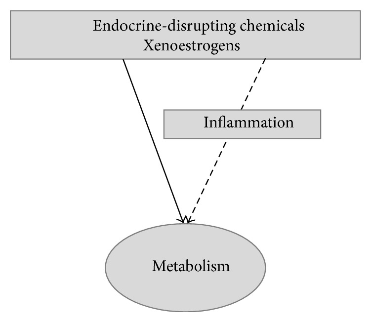
There is extensive evidence supporting the interference of inflammatory activation with metabolism. Obesity, mainly visceral obesity, is associated with a low-grade inflammatory state, triggered by metabolic surplus where specialized metabolic cells such as adipocytes activate cellular stress initiating and sustaining the inflammatory program. The increasing prevalence of obesity, resulting in increased cardiometabolic risk and precipitating illness such as cardiovascular disease, type 2 diabetes, fatty liver, cirrhosis, and certain types of cancer, constitutes a good example of this association. The metabolic actions of estrogens have been studied extensively and there is also accumulating evidence that estrogens influence immune processes. However, the connection between these two fields of estrogen actions has been underacknowledged since little attention has been drawn towards the possible action of estrogens on the modulation of metabolism through their anti-inflammatory properties. In the present paper, we summarize knowledge on the modification inflammatory processes by estrogens with impact on metabolism and highlight major research questions on the field. Understanding the regulation of metabolic inflammation by estrogens may provide the basis for the development of therapeutic strategies to the management of metabolic dysfunctions.


Figures

Figure 1
Figure 1
Estrogen metabolism. The effects of experimental manipulation of enzymes marked in grey boxes modulation are highlighted. Inactivation of aromatase leads to metabolic dysfunction that can be reversed by estrogen replacement. In opposite, estrogen sulfotransferase inactivation in models of obesity or type 2 diabetes mellitus improves metabolic function, an effect that is abolished by ovariectomy. Hepatic expression of steroid sulfatase is induced in animal models of obesity and type 2 diabetes mellitus and seems to alleviate dysmetabolic changes; this effect is also lost with ovariectomy. 17Beta-HSD, 17beta-hydroxysteroid dehydrogenase.
Figure 2
Figure 2
Estrogen signaling occurs through both genomic and nongenomic mechanisms. In classical, genomic, estrogen signaling ERs act as ligand-activated transcription factors, activating or repressing target genes within hours of ligand binding. ERs are located as monomers in the cytoplasm in protein complexes involving heat-shock proteins and estrogen binding promotes their dissociation from this complex and ER dimerization. ER dimers bind directly to estrogen response elements of target gene promoters, or indirectly through interaction with other DNA-bound transcription factors. ERs also regulate gene expression in a ligand independent manner being activated downstream to growth factors binding to growth factor receptors, through the action of intracellular kinases or though the formation of heterodimers with different nuclear receptors (not shown). Genomic actions are modulated by cell-specific interaction with cofactors (coactivators or cosuppressors). Metabolic effects of estrogens seem to be largely mediated through nonnuclear ERs, either by interference with gene expression or by exerting nongenomic actions. This involves activation of ERs and G-protein-coupled ER located at the membrane or at extranuclear sites within seconds or minutes resulting in changes in Ca2+, K+, cAMP, and nitric oxide levels, activation of G protein-mediated events, and stimulation of different types of kinases such as extracellular-regulated kinases, phosphoinositide 3-kinases, mitogen-activated protein kinase, and c-Jun N-terminal kinases. E: estrogen; ER: estrogen receptor; ERE: estrogen-responsive element; ERK: extracellular-regulated kinase; GFR: growth factor receptor; GPER: G protein-coupled estrogen receptor; HSP: heat-shock protein; JNK: c-Jun N-terminal kinase; MAPK: mitogen-activated protein kinase; NO: nitric oxide; PI3K: phosphoinositide-3 kinase; TF: transcription factor.
Figure 3
Figure 3
Different models have shown the influence of estrogens on metabolic-related inflammation. Loss of/decreased estrogen signaling through decreased production of estrogens or ERalpha, ERalpha/beta, or GPER inactivation promotes metabolic dysfunction revealed by visceral obesity, insulin resistance, dyslipidemia, inflammatory activation, and nonalcoholic fatty liver disease. On the other hand, promoting maintenance of estrogen signaling through hormone replacement therapy, blocking estrogen inactivation by estrogen sulfotransferase or increasing its reactivation from the estrogen-sulfate circulating pool by steroid sulfatase induction, tends to counteract metabolic dysfunction. Interestingly, inactivation of ERbeta also promotes metabolic health, showing the opposite metabolic effects mediated by both ER receptors. ER: estrogen receptor; GPER: G protein-coupled estrogen receptor; HRT: hormone replacement therapy; NAFLD: nonalcoholic fatty liver disease.
Figure 4
Figure 4
Estrogens effects on metabolic improvement may be cause (a) or consequence (b) of regulation of inflammation pathways. Further studies are needed to ascertain the relationship between estrogen signaling, inflammation, and metabolism. It is possible that the anti-inflammatory effects of estrogens, though their influences on processes like energy balance, leptin and glucocorticoid signaling, adipose tissue distribution and cellularity, and activity of immune cells, may culminate on metabolic improvement. However, estrogens have also been demonstrated to directly interfere with such processes, favoring improved inflammatory profile that results in overall metabolic amelioration. Additionally, the two hypotheses are not mutually exclusive. AT: adipose tissue.
Figure 5
Figure 5
Effects of xenoestrogens on inflammation may mediate their actions on metabolism.

References

    1. Hotamisligil G. S. Inflammation and metabolic disorders. Nature. 2006;444(7121):860–867. doi: 10.1038/nature05485. - DOI - PubMed
    1. Mathis D., Shoelson S. E. Immunometabolism: an emerging frontier. Nature Reviews Immunology. 2011;11(2):81–83. doi: 10.1038/nri2922. - DOI - PMC - PubMed
    1. Gregor M. F., Hotamisligil G. S. Inflammatory mechanisms in obesity. Annual Review of Immunology. 2011;29:415–445. doi: 10.1146/annurev-immunol-031210-101322. - DOI - PubMed
    1. Schäffler A., Schölmerich J., Salzberger B. Adipose tissue as an immunological organ: toll-like receptors, C1q/TNFs and CTRPs. Trends in Immunology. 2007;28(9):393–399. doi: 10.1016/j.it.2007.07.003. - DOI - PubMed
    1. Monteiro R. Chronic inflammation in the metabolic syndrome—emphasis on adipose tissue. In: Soares R., Costa C., editors. Oxidative Stress, Inflammation and Angiogenesis in the Metabolic Syndrome. London, UK: Springer; 2009. pp. 65–83.

Publication types

LinkOut - more resources