

18F-fluorodeoxyglucose positron emission tomography/computed tomography in a case of non-ketotic hyperglycemia
- PMID: 25400367
- PMCID: PMC4228591
- DOI: 10.4103/0972-3919.142633
18F-fluorodeoxyglucose positron emission tomography/computed tomography in a case of non-ketotic hyperglycemia
Abstract
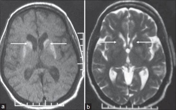
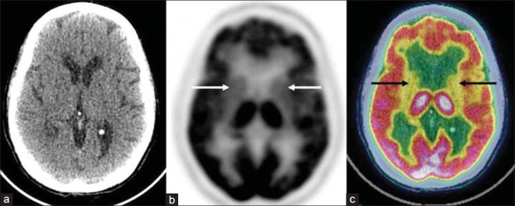
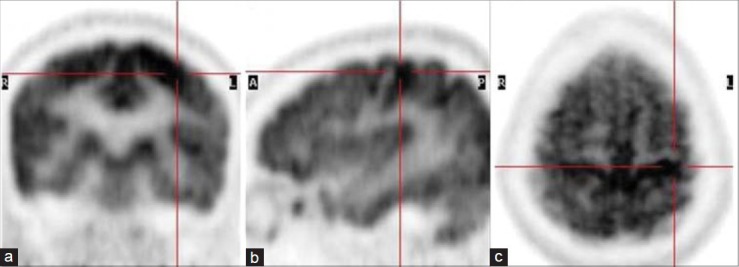
Hemichorea and generalized chorea are rare syndromes associated with nonketotic hyperglycemia. This disorder usually afflicts elderly females, and may herald the onset of new onset diabetes, usually type 2. There are conflicting reports of the underlying pathophysiology of this rare entity. Magnetic resonance imaging findings have been described in the past, and are characteristic. There are very few reports of 18F-fluorodeoxyglucose positron emission tomography/computed tomography (PET/CT) findings of this unusual dyskinetic syndrome. This report describes the PET/CT features of this rare disease. Early detection and prompt correction of hyperglycemia may lead to complete or significant amelioration of symptoms.
Keywords: 18F-fluorodeoxyglucose positron emission tomography/computed tomography; chorea; nonketotic hyperglycemia.
Conflict of interest statement
Figures
References
-
- Bidwell SF. Some observations on hemballismus. Neurol. 1960;10:619–26.
-
- Lin JJ, Lin GY, Shih C, Shen WC. Presentation of striatal hyperintensity on T1-weighted MRI in patients with hemiballism-hemichorea caused by non-ketotic hyperglycemia: Report of seven new cases and a review of literature. J Neurol. 2001;248:750–5. - PubMed
-
- Yacoub HA. Abnormal magnetic resonance imaging and hemichorea associated with non-ketotic hyperglycemia. J Neurol Res. 2013;3:146–9.
-
- Hsu JL, Wang HC, Hsu WC. Hyperglycemia-induced unilateral basal ganglion lesions with and without hemichorea. A PET study. J Neurol. 2004;251:1486–90. - PubMed
-
- Nguyen BD. Brain and upper extremity PET/CT findings of hyperglycemia-induced hemiballism-hemichorea. Clin Nucl Med. 2007;32:643–5. - PubMed
Publication types
LinkOut - more resources
Full Text Sources
Other Literature Sources
Miscellaneous
