

Magnifying endoscopy of gastric epithelial dysplasia based on the morphologic characteristics
- PMID: 25400462
- PMCID: PMC4229543
- DOI: 10.3748/wjg.v20.i42.15771
Magnifying endoscopy of gastric epithelial dysplasia based on the morphologic characteristics
Abstract
Aim: To investigate the difference in magnifying endoscopic findings of gastric epithelial dysplasias (GEDs) according to the morphologic characteristics.
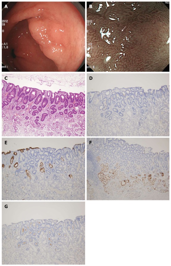
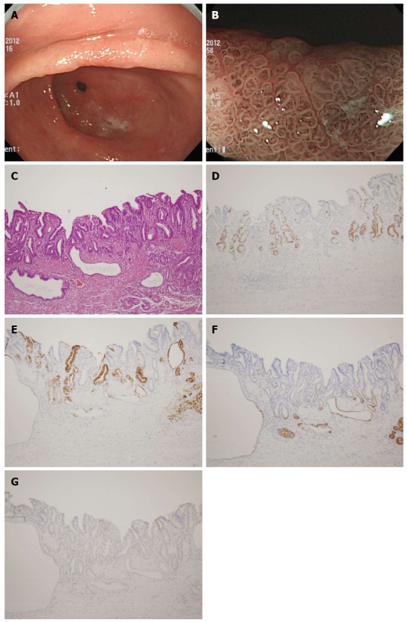
Methods: This study included 46 GED lesions in 45 patients who underwent magnifying endoscopy using narrow band imaging (ME-NBI) before endoscopic resection. During ME-NBI, the microvascular and microsurface (MS) patterns and the presence of light blue crest (LBC) and white opaque substance were investigated. GEDs were categorized as adenomatous, foveolar, and hybrid types, and their mucin phenotype was evaluated.
Results: Of the 46 lesions, 27 (59%) were categorized as adenomatous, 15 (32%) as hybrid, and the remaining 4 (9%) as foveolar. All adenomatous GEDs showed the round pit and/or tubular MS patterns, all foveolar GEDs showed the papillary pattern, and hybrid GEDs showed mixed patterns (P < 0.001). LBC was more frequently observed in adenomatous GEDs than in hybrid or foveolar GEDs (52%, 33%, 0%, respectively), although this difference was not significant (P = 0.127). The papillary MS pattern was associated with MUC5AC and MUC6 expression, and the round pit and/or tubular MS patterns were associated with CD10 expression.
Conclusion: The MS pattern in ME-NBI findings is useful for predicting the morphologic category and mucin phenotype of GEDs, and ME-NBI findings may guide decisions regarding GED treatment.
Keywords: Dysplasia; Magnifying endoscopy; Mucin; Narrow band imaging; Stomach.
Figures
Similar articles
-
Adenomatous and foveolar gastric dysplasia: distinct patterns of mucin expression and background intestinal metaplasia.Am J Surg Pathol. 2008 Apr;32(4):524-33. doi: 10.1097/PAS.0b013e31815b890e. Am J Surg Pathol. 2008. PMID: 18300795
-
Gastric epithelial dysplasia: characteristics and long-term follow-up results after endoscopic resection according to morphological categorization.BMC Gastroenterol. 2015 Feb 12;15:17. doi: 10.1186/s12876-015-0249-7. BMC Gastroenterol. 2015. PMID: 25886985 Free PMC article.
-
Magnifying Endoscopy with Narrow Band Imaging of Early Gastric Cancer: Correlation with Histopathology and Mucin Phenotype.Gut Liver. 2016 Jul 15;10(4):532-41. doi: 10.5009/gnl15364. Gut Liver. 2016. PMID: 27021504 Free PMC article.
-
Usefulness of magnifying narrow-band imaging endoscopy for the diagnosis of gastric and colorectal lesions.Digestion. 2012;85(2):74-9. doi: 10.1159/000334642. Epub 2012 Jan 19. Digestion. 2012. PMID: 22269282 Review.
-
Diagnostic performance of magnifying narrow-band imaging for early gastric cancer: A meta-analysis.World J Gastroenterol. 2015 Jul 7;21(25):7884-94. doi: 10.3748/wjg.v21.i25.7884. World J Gastroenterol. 2015. PMID: 26167089 Free PMC article. Review.
Cited by
-
Efforts to increase image quality during endoscopy: The role of pronase.World J Gastrointest Endosc. 2016 Mar 10;8(5):267-72. doi: 10.4253/wjge.v8.i5.267. World J Gastrointest Endosc. 2016. PMID: 26981178 Free PMC article. Review.
-
Endoscopic Submucosal Dissection of Gastric High-Grade Foveolar Dysplasia With Normal Background Mucosa.Gastro Hep Adv. 2024 Oct 10;4(2):100565. doi: 10.1016/j.gastha.2024.10.005. eCollection 2025. Gastro Hep Adv. 2024. PMID: 39866717 Free PMC article.
References
-
- Lewin KJ, Appleman HD. Carcinoma of the stomach. In: Rosai J ed., editor. Atlas of Tumor Pathology. Tumors of the Esophagus and Stomach (3rd Series Fascicle 18) Washington, DC: AFIP; 1996. pp. 245–330.
-
- Lauwers GY. Defining the pathologic diagnosis of metaplasia, atrophy, dysplasia, and gastric adenocarcinoma. J Clin Gastroenterol. 2003;36:S37–43; discussion S61-2. - PubMed
-
- Abraham SC, Montgomery EA, Singh VK, Yardley JH, Wu TT. Gastric adenomas: intestinal-type and gastric-type adenomas differ in the risk of adenocarcinoma and presence of background mucosal pathology. Am J Surg Pathol. 2002;26:1276–1285. - PubMed
-
- Abraham SC, Park SJ, Lee JH, Mugartegui L, Wu TT. Genetic alterations in gastric adenomas of intestinal and foveolar phenotypes. Mod Pathol. 2003;16:786–795. - PubMed
Publication types
MeSH terms
Substances
LinkOut - more resources
Full Text Sources
Other Literature Sources
Medical
