

Clinicopathological and immunohistochemical features of primary central nervous system germ cell tumors: a 24-years experience
- PMID: 25400782
- PMCID: PMC4230147
Clinicopathological and immunohistochemical features of primary central nervous system germ cell tumors: a 24-years experience
Abstract
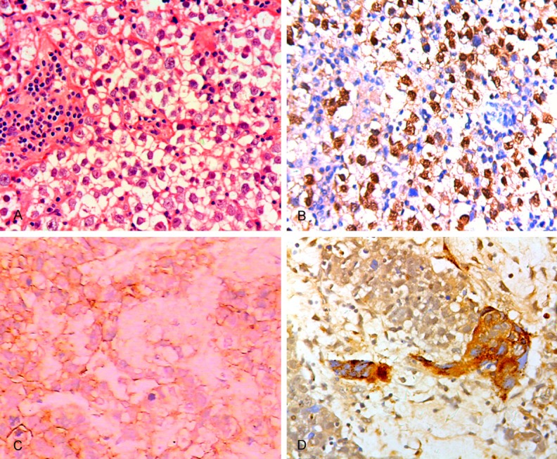
Primary central nervous system (CNS) germ cell tumors (GCTs) are a rare heterogeneous group of lesions, which the clinicopathological features have a marked degree of heterogeneity comparing with that of gonadal GCTs. Accurately diagnosing CNS GCTs might be extremely difficult and requires immunohistochemical verification. This study was to investigate the biological feature of CNS GCTs and diagnostic value of immunohistochemical markers OCT3/4, C-kit, PLAP, and CD30 in CNS GCTs. A retrospective study was performed on 34 patients with CNS germ cell tumors between 1990 and 2014. 34 CNS GCTs account for 9.2% of all primary CNS neoplasms. The sellar region (35.3%) and pineal gland (17.6%) were the most common sites of intracranial GCTs. Hydrocephalus (82.4%) and diplopia (46.9%) were the two most common clinical presentations. The most common histological subtypes were germinoma (67.6%). PLAP, c-kit, OCT3/4 were highly expressed in gernimomas. CD30 and CK AE1/3 stainings were positive in embryonal carcinoma. Yolk sac tumor component showed positive staining for AFP and CK AE1/3. β-HCG staining was positive in choriocarcinoma and STGC. Patients with mature teratomas and germinomas had a better prognosis (a 5-year survival rate) than those with embryonal carcinoma and choriocarcinoma (a 5-year survival rates were 0). Our finding suggest that the incidences of primary CNS GCTs are higher in South China than in the West, but mixed GCTs are uncommon in our study. The judicious use of a panel of selected markers is helpful in diagnosing and predicting the prognosis for CNS GCTs.
Keywords: C-kit; CD30; Central nervous system; OCT3/4; PLAP; germ cell tumors.
Figures
References
-
- Thakkar JP, Chew L, Villano JL. Primary CNS germ cell tumors: current epidemiology and update on treatment. Med Oncol. 2013;30:496–515. - PubMed
-
- Packer RJ, Cohen BH, Cooney K. Intracranial germ cell tumors. Oncologist. 2000;5:312–320. - PubMed
-
- Lee D, Suh YL. Histologically confirmed intracranial germ cell tumors; an analysis of 62 patients in a single institute. Virchows Arch. 2010;457:347–357. - PubMed
-
- Jennings MT, Gelman R, Hochberg F. Intracranial germ-cell tumors: natural history and pathogenesis. J Neurosurg. 1985;63:155–167. - PubMed
Publication types
MeSH terms
Substances
LinkOut - more resources
Full Text Sources
Research Materials
Miscellaneous