

Clinicopathological findings of primary esophageal malignant melanoma: report of six cases and review of literature
- PMID: 25400820
- PMCID: PMC4230078
Clinicopathological findings of primary esophageal malignant melanoma: report of six cases and review of literature
Abstract
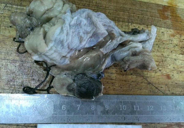
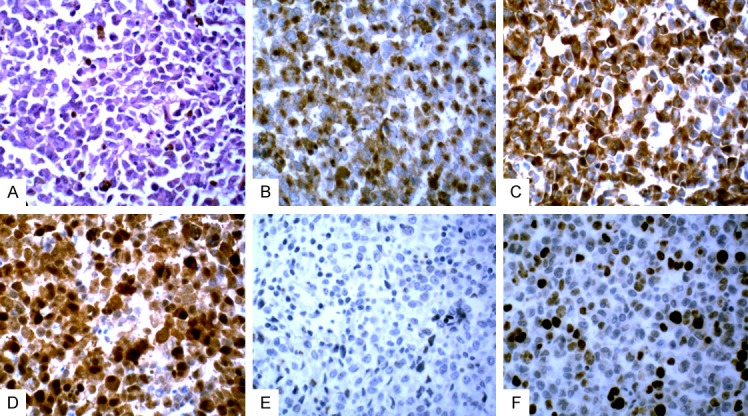
We studied images and histopathological features of primary esophageal malignant melanoma to explore the clinical pathological features, diagnosis, differential diagnoses, and treatment. Immunolabelling was conducted on six cases of esophageal malignant melanoma using histological and immunohistochemical techniques. Combined with the related literature, the clinical manifestations, imaging, histopathological and immunohistochemical features, treatment, and prognosis of primary esophageal malignant melanoma were observed and analyzed. The six patients with primary esophageal malignant melanoma were all male with an average age of 63.4 years. Poor food intake was observed in all patients, and the symptoms showed progressive aggravation. Endoscopic feed tube revealed dark brown and black nodular and polypoid lesions, 1/4-1/2 loop cavity. Tumor histopathology revealed the following characteristics: tumor cells arranged in nests, sheets and cords, round or polygonal, abundant and red-stained cytoplasm, melanin granules in the cytoplasm, heterogeneous nucleus sizes, centered or deviated nuclei, clearly identifiable nucleoli, and apparent pathological mitosis. The immune phenotype was as follows: tumor cells had diffuse expression of HMB45, Melan A, and S100. The cells were CK negative, and the Ki67-positive cell number was 40%-45%. Primary esophageal malignant melanoma is rare with high malignancy and poor prognosis. Immunohistochemical staining is helpful for diagnosing this tumor. The differential diagnosis includes low differentiated carcinoma, primitive neuroectodermal tumor, esophageal sarcomatoid carcinoma, esophageal lymphoma, and other tumors.
Keywords: Esophagus; differential diagnosis; immunohistochemistry; malignant melanoma; tumor.
Figures

Similar articles
-
Difficulties encountered in the diagnosis of primary esophageal malignant melanoma by 18F-fluorodeoxyglucose positron emission tomography/computed tomography: a case report.Ann Palliat Med. 2021 Apr;10(4):4975-4981. doi: 10.21037/apm-21-649. Ann Palliat Med. 2021. PMID: 33966432
-
Clinicopathological features of primary hepatic diffuse large B-cell lymphoma: a report of seven cases and a literature review.Int J Clin Exp Pathol. 2015 Oct 1;8(10):12955-60. eCollection 2015. Int J Clin Exp Pathol. 2015. PMID: 26722490 Free PMC article. Review.
-
Primary malignant melanoma of the esophagus: a case report.World J Gastroenterol. 2014 Mar 14;20(10):2731-4. doi: 10.3748/wjg.v20.i10.2731. World J Gastroenterol. 2014. PMID: 24627611 Free PMC article.
-
Esophageal subepithelial lesion diagnosed as malignant gastrointestinal neuroectodermal tumor.World J Gastroenterol. 2015 May 14;21(18):5739-43. doi: 10.3748/wjg.v21.i18.5739. World J Gastroenterol. 2015. PMID: 25987801 Free PMC article. Review.
-
[Primary esophageal melanoma].Gastroenterol Hepatol. 1998 Jun-Jul;21(6):283-6. Gastroenterol Hepatol. 1998. PMID: 9711011 Spanish.
Cited by
-
The Thousand Faces of Malignant Melanoma: A Systematic Review of the Primary Malignant Melanoma of the Esophagus.Cancers (Basel). 2022 Jul 30;14(15):3725. doi: 10.3390/cancers14153725. Cancers (Basel). 2022. PMID: 35954389 Free PMC article. Review.
-
Clinicopathological characteristics and survival of primary malignant melanoma of the esophagus.Oncol Lett. 2019 Aug;18(2):1872-1880. doi: 10.3892/ol.2019.10519. Epub 2019 Jun 21. Oncol Lett. 2019. PMID: 31423256 Free PMC article.
-
Clinicopathological characterization of ten patients with primary malignant melanoma of the esophagus and literature review.World J Gastrointest Oncol. 2022 Sep 15;14(9):1739-1757. doi: 10.4251/wjgo.v14.i9.1739. World J Gastrointest Oncol. 2022. PMID: 36187400 Free PMC article.
-
How to update esophageal masses imaging using literature review (MRI and CT features).Insights Imaging. 2024 Jul 6;15(1):169. doi: 10.1186/s13244-024-01754-0. Insights Imaging. 2024. PMID: 38971944 Free PMC article. Review.
-
Primary malignant melanoma of esophagus following chemoradiotherapy for esophageal squamous cell carcinoma: report of a case.Clin J Gastroenterol. 2017 Aug;10(4):336-341. doi: 10.1007/s12328-017-0751-2. Epub 2017 May 26. Clin J Gastroenterol. 2017. PMID: 28550655
References
-
- Oshiro T, Shimoji H, Mstsuura F, Uchima N, Kinjo F, Nakayama T, Nishimaki T. Primary malignant melanoma of the esophagus arising from a melanotic lesion: report of a case. Surg Today. 2007;37:671–675. - PubMed
-
- Lin PW, Lee RC, Chern MS, Chiang JH, Chang CY. Primary malignant melanoma of the esophagus. J Chin Med Assoc. 2006;69:334–337. - PubMed
-
- Li B, Lei W, Shao K, Zhang C, Chen Z, Shi S, He J. Characteristics and prognosis of primary malignant melanoma of the esophagus. Melanoma Res. 2007;17:239–242. - PubMed
Publication types
MeSH terms
Substances
LinkOut - more resources
Full Text Sources
Medical
Research Materials