. 2014 Sep 15;7(10):7249-61.
eCollection 2014.

MiR-378 overexpression attenuates high glucose-suppressed osteogenic differentiation through targeting CASP3 and activating PI3K/Akt signaling pathway

Affiliations

MiR-378 overexpression attenuates high glucose-suppressed osteogenic differentiation through targeting CASP3 and activating PI3K/Akt signaling pathway

Li You et al. Int J Clin Exp Pathol. .

Abstract

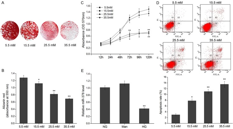
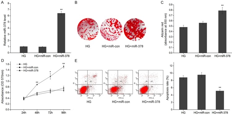
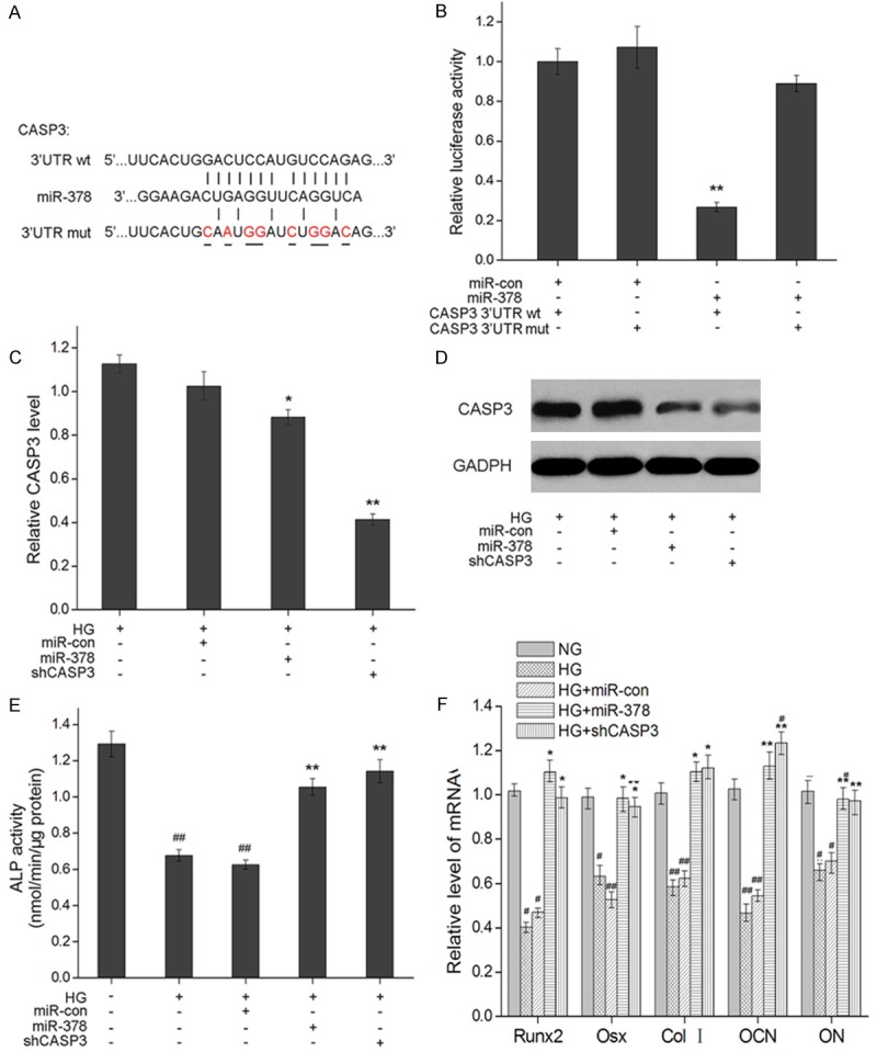
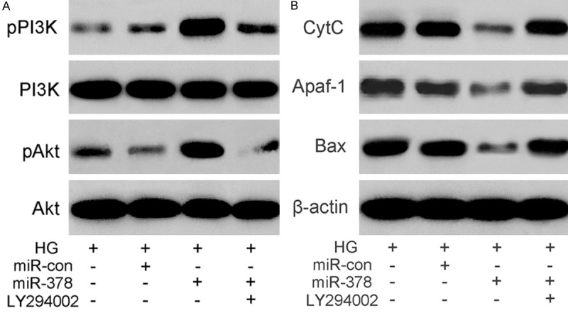
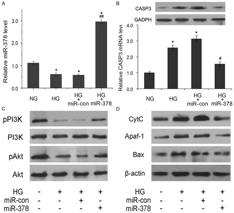
Hyperglycemia is one of the possible causes for osteoporosis and bone fracture in diabetes mellitus. Here we modeled diabetes-induced osteoporosis in vitro using preosteoblastic cell line MC3T3-E1 and a diabetic mice model for in vivo studies. We found that in addition to reducing osteoblast viability and differentiation (mineralization), culture in elevated glucose down regulated microRNA-378 (miR-378) expression but ectopic miR-378 expression reversed the effects of high glucose. We identified caspase-3 (CASP3) as a target of miR-378 and showed that miR-378 repressed CASP3 mRNA and protein expression under high glucose condition. We further showed that both miR-378 expression and CASP3 silencing independently restored alkaline phosphatase (ALP) activity and the expression of osteoblastic differentiation markers Runt-related transcription factor 2 (Runx2), osteorix (Osx), collagen I (Col I), osteocalcin (OCN), and osteonectin (ON). We also found that under high glucose conditions miR-378 activated the PI3K/Akt signaling pathway and down regulated pro-apoptotic CytC, Apaf-1 and Bax proteins via the PI3K/Akt pathway. Collectively, these results suggest that miR-378 overexpression attenuates high glucose-suppressed osteogenic differentiation through targeting CASP3 and activating the PI3K/Akt pathway.

Keywords: CASP3; MiR-378; PI3K/Akt signaling; high glucose; osteogenic differentiation.


Figures

Figure 1
Figure 1
High glucose inhibits cell growth and mineralization of MC3T3-E1 and down regulates miR-378. A. Mineralization of osteoblasts cultured in glucose (5.5-35.5 mM) for 21 d depicted by Alizarin Red S staining. B. Alizarin Red S quantification by absorbance (at 550 nm) of osteoblasts cultured in Glucose (5.5-35.5 mM) for 21 d. C. Viability of osteoblasts cultured in glucose (5.5-35.5 mM) for 12-120 h represented by production of formazan in an MTT assay. D. Cell apoptosis of MC3T3-E1 cells after incubation with the indicated concentration of glucose for 120 h was evaluated by flow cytometry using Annexin V/PI-double staining. The apoptosis rates were the values of Q2 quadrant (Annexin V+/PI+) plus Q4 quadrant (Annexin V+/PI-). E. Relative miR-378 levels assayed by qRT-PCR in MC3T3-E1 cells cultured in no glucose (NG), mannose (Man) or high glucose (HG, 25.5 mM) for 120 h. *P < 0.05, **P < 0.01.
Figure 2
Figure 2
MiR-378 overexpression improves cell viability and mineralization under high glucose. A. Relative qRT-PCR expression levels of miR-378 48 h after transfection in MC3T3-E1, MC3T3-E1/miR-con and MC3T3-E1/miR-378 cells under high glucose (HG, 25.5 mM). B. Mineralization of osteoblasts cultured in HG for 21 d depicted by Alizarin Red S staining. C. Alizarin Red S quantification by absorbance (at 550 nm) of osteoblasts cultured in HG for 21 d. D. Viability of MC3T3-E1, MC3T3-E1/miR-con and MC3T3-E1/miR-378 cells cultured in HG for 24-96 h represented by production of formazan in an MTT assay. E. Representative flow cytometric analysis of apoptosis in MC3T3-E1 cells 48 h after transfection. The apoptosis rates were the values of Q2 quadrant plus Q4 quadrant. *P < 0.05, **P < 0.01.
Figure 3
Figure 3
MiR-378 post transcriptionally down-regulates CASP3 expression by targeting its 3’UTR. A. Sequence complementarity between mmu-miR-378 and the seed region in the CASP3 3’UTR. Base pair mutations in the mutation-bearing construct are underlined. Short vertical lines indicate complementary nucleotides. B. Relative luciferase activity in MC3T3-E1 cells cultured in high glucose (HG, 25 mM) 24 h after co-transfection of miR-378 (Ambion) or mock vector (miR-con) with wild-type (wt) or mutant (mut) CASP3 3’UTR-luciferase reporter vector. Measurements from three biological replicates for each combination of constructs are shown along with SEM and **indicate P < 0.01. C. CASP3 mRNA expression levels 48 h after transfection in MC3T3-E1, MC3T3-E1/miR-con and MC3T3-E1/miR-378 and MC3T3-E1/shCASP3 cells cultured in HG. Relative mRNA expression was detected by qRT-RCR. GADPH was used as an internal control. D. Western blot assays of CASP3 protein levels 48 h after transfection in MC3T3-E1, MC3T3-E1/miR-con and MC3T3-E1/miR-378 and MC3T3-E1/shCASP3 cells cultured under HG. E. ALP activity of MC3T3-E1, MC3T3-E1/miR-con and MC3T3-E1/miR-378 and MC3T3-E1/shCASP3 cells cultured in high glucose (25.5 mM) for 14 days. *P < 0.05 and **P < 0.01 compared to MC3T3-E1 cells cultured in HG; #P < 0.05 and ##P < 0.01 compared to MC3T3-E1 cells cultured in 5.5 mM glucose. F. mRNA expression levels of osteogenic marker genes in MC3T3-E1, MC3T3-E1/miR-con, MC3T3-E1/miR-378 and MC3T3-E1/shCASP3 cells cultured in HG for 2 days *P < 0.05 and **P < 0.01 compared with MC3T3-E1 in HG; #P < 0.05 and ##P < 0.01 compared with MC3T3-E1 cells in physiological levels of glucose (NG).
Figure 4
Figure 4
MiR-378 down regulates expression of apoptosis-related proteins through the activation of the PI3K/Akt pathway. (A) The expression of p-PI3K, p-Akt and (B) apoptosis proteins Cyt C, Apaf-1, and Bax detected by western blotting of protein samples from high glucose (25.5 mM) cultures of MC3T3-E1/miR-378 cells treated (+) or not treated (-) with PI3K inhibitor LY294002. β-actin was an internal control.
Figure 5
Figure 5
MiR-378 regulates expression of CASP3 and PI3K/Akt pathway in diabetic model mice. (A) Relative expression levels of miR-378 in tibias of normal and diabetic mice injected with miR-con (HG+miR-con group) or miR-378 (HG+miR-378 group). (B) Relative mRNA and protein expressions of CASP3 in rat tibias. (C) The expression of p-PI3K, p-Akt and apoptosis proteins Cyt C, Apaf-1, and Bax (D) detected by western blotting of protein samples from tibias of normal and diabetic mice injected with miR-con or miR-378. β-actin was an internal control. *P < 0.05 compared with normal mice (NG group); #P < 0.05 and ##P < 0.01 compared with diabetic mice (HG group).

References

    1. Blakytny R, Spraul M, Jude EB. Review: The diabetic bone: a cellular and molecular perspective. Int J Low Extrem Wounds. 2011;10:16–32. - PubMed
    1. Bouillon R. Diabetic bone disease. Calcif Tissue Int. 1991;49:155–160. - PubMed
    1. Rakel A, Sheehy O, Rahme E, LeLorier J. Osteoporosis among patients with type 1 and type 2 diabetes. Diabetes Metab. 2008;34:193–205. - PubMed
    1. Merlotti D, Gennari L, Dotta F, Lauro D, Nuti R. Mechanisms of impaired bone strength in type 1 and 2 diabetes. Nutr Metab Cardiovasc Dis. 2010;20:683–690. - PubMed
    1. Balint E, Szabo P, Marshall CF, Sprague SM. Glucose-induced inhibition of in vitro bone mineralization. Bone. 2001;28:21–28. - PubMed

MeSH terms

LinkOut - more resources