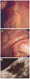
Recto-sigmoid endoscopic-ultrasonography in the staging of deep infiltrating endometriosis
- PMID: 25400866
- PMCID: PMC4231491
- DOI: 10.4253/wjge.v6.i11.525
Recto-sigmoid endoscopic-ultrasonography in the staging of deep infiltrating endometriosis
Abstract
Recto-sigmoid endoscopic ultrasonography (RS-EUS) has first been used in the staging of pelvic deep infiltrating endometriosis in the early 1990's. Since then, although publications have been sparse, RS-EUS is routinely used for this indication in few centers. In this paper, we focus on technical aspects and operating method of rectal and sigmoid endo-sonography, and describe the most characteristic echographic presentations of endometriosis of the lower digestive tract. Through a literature review, results obtained with different types of endo-rectal probes, either flexible endoscopic, or blind rigid, are presented and compared with those of other close imaging techniques: magnetic resonance imaging and the more recent trans-vaginal sonography. As well as these two latter techniques, RS-EUS appears as an interesting method in the staging of pelvic deep infiltrating endometriosis particularly to evaluate rectal and sigmoid infiltrations. However, more prospective studies are required, to correctly define respective indications for each exam, in the light of recent advancements in treating this frequent disease.
Keywords: Endometriosis; Endoscopic-ultrasonography; Magnetic resonance imaging; Rectum and sigmoid; Surgical treatment; Ultrasound.
Figures


Similar articles
-
Comparison of endoscopic ultrasound and magnetic resonance imaging in severe pelvic endometriosis.Gastroenterol Clin Biol. 2000 Dec;24(12):1197-204. Gastroenterol Clin Biol. 2000. PMID: 11173733 English, French.
-
Rectosigmoid endometriosis: endoscopic ultrasound features and clinical implications.Endoscopy. 2000 Jul;32(7):525-30. doi: 10.1055/s-2000-9008. Endoscopy. 2000. PMID: 10917184
-
Deep infiltrating endometriosis: Should rectal and vaginal opacification be systematically used in MR imaging?Gynecol Obstet Fertil. 2016 Jun;44(6):322-8. doi: 10.1016/j.gyobfe.2016.03.016. Epub 2016 May 20. Gynecol Obstet Fertil. 2016. PMID: 27216959
-
Deep endometriosis infiltrating the recto-sigmoid: critical factors to consider before management.Hum Reprod Update. 2015 May-Jun;21(3):329-39. doi: 10.1093/humupd/dmv003. Epub 2015 Jan 24. Hum Reprod Update. 2015. PMID: 25618908 Review.
-
Imaging Modalities for Diagnosis of Deep Pelvic Endometriosis: Comparison between Trans-Vaginal Sonography, Rectal Endoscopy Sonography and Magnetic Resonance Imaging. A Head-to-Head Meta-Analysis.Diagnostics (Basel). 2019 Dec 17;9(4):225. doi: 10.3390/diagnostics9040225. Diagnostics (Basel). 2019. PMID: 31861142 Free PMC article. Review.
Cited by
-
Preoperative rectosigmoid endoscopic ultrasonography predicts the need for bowel resection in endometriosis.World J Gastroenterol. 2019 Feb 14;25(6):696-706. doi: 10.3748/wjg.v25.i6.696. World J Gastroenterol. 2019. PMID: 30783373 Free PMC article.
-
Diagnostic value of endoscopic ultrasound in pelvic masses with bowel involvement.Int J Surg. 2024 Apr 1;110(4):2085-2091. doi: 10.1097/JS9.0000000000001124. Int J Surg. 2024. PMID: 38668660 Free PMC article.
-
Outcomes of discoid excision and segmental resection for colorectal endometriosis: robotic versus conventional laparoscopy.J Robot Surg. 2024 Feb 22;18(1):87. doi: 10.1007/s11701-024-01854-5. J Robot Surg. 2024. PMID: 38386205
-
Changes in hospital consumption of opioid and non-opioid analgesics after colorectal endometriosis surgery.J Robot Surg. 2023 Dec;17(6):2703-2710. doi: 10.1007/s11701-023-01691-y. Epub 2023 Aug 22. J Robot Surg. 2023. PMID: 37606871
-
Rectal Endoscopic Ultrasound in Clinical Practice.Curr Gastroenterol Rep. 2019 Apr 12;21(4):18. doi: 10.1007/s11894-019-0682-9. Curr Gastroenterol Rep. 2019. PMID: 30980194 Review.
References
-
- Chapron C, Fauconnier A, Dubuisson JB, Barakat H, Vieira M, Bréart G. Deep infiltrating endometriosis: relation between severity of dysmenorrhoea and extent of disease. Hum Reprod. 2003;18:760–766. - PubMed
-
- Koninckx PR, Martin DC. Deep endometriosis: a consequence of infiltration or retraction or possibly adenomyosis externa? Fertil Steril. 1992;58:924–928. - PubMed
-
- Zwas FR, Lyon DT. Endometriosis. An important condition in clinical gastroenterology. Dig Dis Sci. 1991;36:353–364. - PubMed
-
- Yantiss RK, Clement PB, Young RH. Endometriosis of the intestinal tract: a study of 44 cases of a disease that may cause diverse challenges in clinical and pathologic evaluation. Am J Surg Pathol. 2001;25:445–454. - PubMed
-
- Chapron C, Dubuisson JB, Chopin N, Foulot H, Jacob S, Vieira M, Barakat H, Fauconnier A. [Deep pelvic endometriosis: management and proposal for a “surgical classification”] Gynecol Obstet Fertil. 2003;31:197–206. - PubMed
Publication types
LinkOut - more resources
Full Text Sources
Other Literature Sources
Research Materials

