. 2013 Oct;1(1):47-62.
doi: 10.1002/iid3.9. Epub 2013 Oct 30.

Relative contribution of IL-1α, IL-1β and TNF to the host response to Mycobacterium tuberculosis and attenuated M. bovis BCG

Affiliations

Relative contribution of IL-1α, IL-1β and TNF to the host response to Mycobacterium tuberculosis and attenuated M. bovis BCG

Marie-Laure Bourigault et al. Immun Inflamm Dis. 2013 Oct.

Abstract

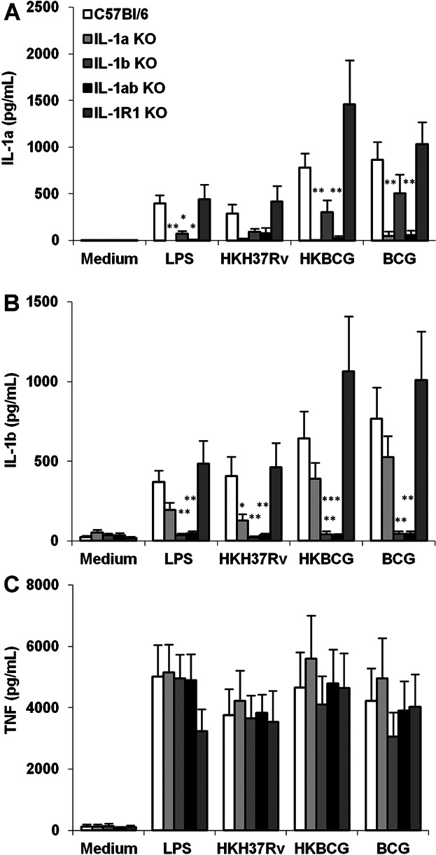
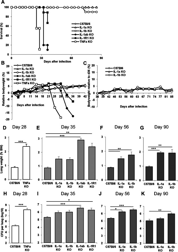
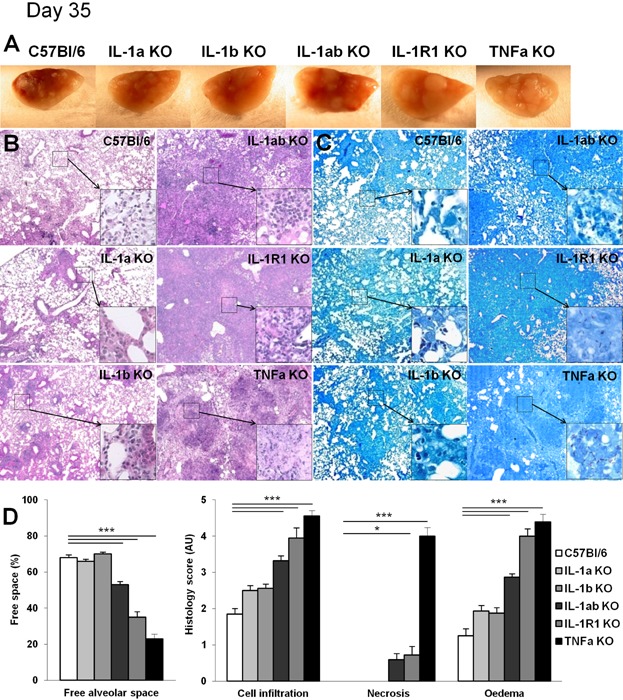
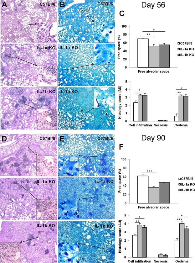
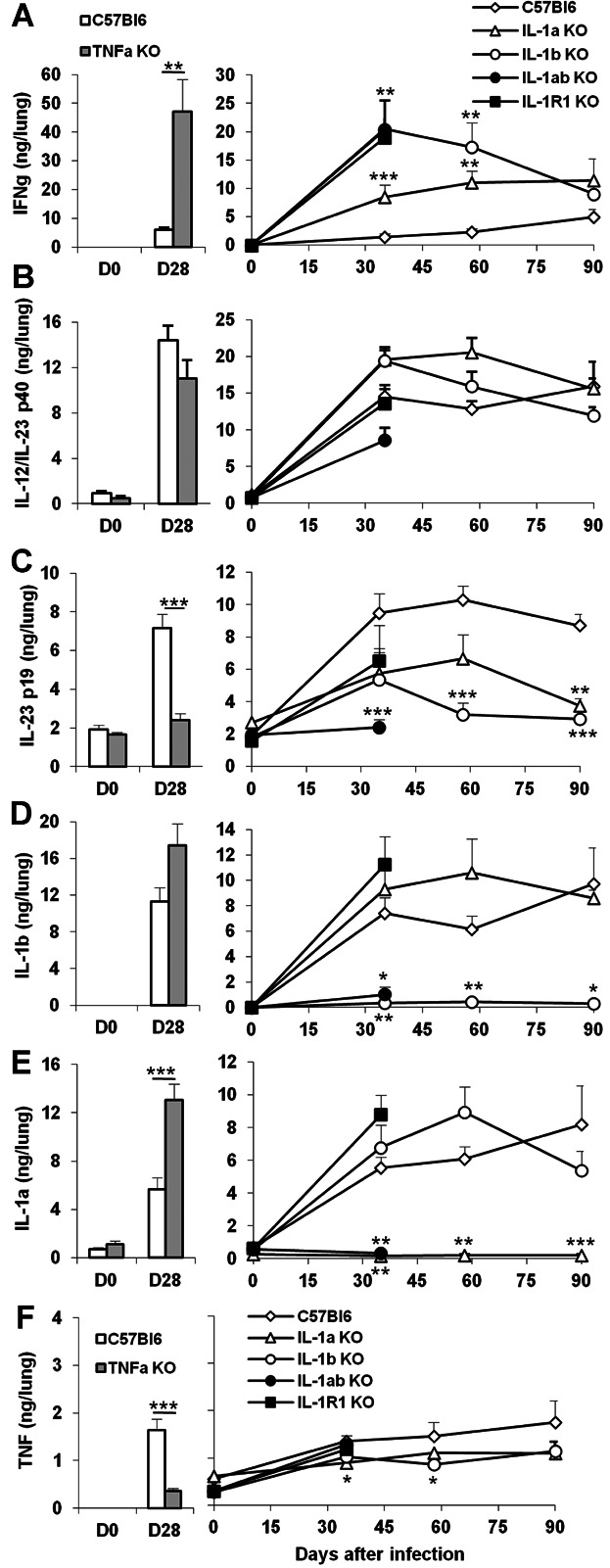
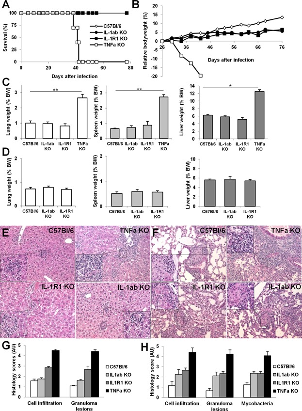
TNF and IL-1 are major mediators involved in severe inflammatory diseases against which therapeutic neutralizing antibodies are developed. However, both TNF and IL-1 receptor pathways are essential for the control of Mycobacterium tuberculosis infection, and it is critical to assess the respective role of IL-1α, IL-1β, and TNF. Using gene-targeted mice we show that absence of both IL-1α and IL-1β recapitulates the uncontrolled M. tuberculosis infection with increased bacterial burden, exacerbated lung inflammation, high IFNγ, reduced IL-23 p19 and rapid death seen in IL-1R1-deficient mice. However, presence of either IL-1α or IL-1β in single-deficient mice is sufficient to control acute M. tuberculosis infection, with restrained bacterial burden and lung pathology, in conditions where TNF deficient mice succumbed within 4 weeks with overwhelming infection. Systemic infection by attenuated M. bovis BCG was controlled in the absence of functional IL-1 pathway, but not in the absence of TNF. Therefore, although both IL-1α and IL-1β are required for a full host response to virulent M. tuberculosis, the presence of either IL-1α or IL-1β allows some control of acute M. tuberculosis infection, and IL-1 pathway is dispensable for controlling M. bovis BCG acute infection. This is in sharp contrast with TNF, which is essential for host response to both attenuated and virulent mycobacteria and may have implications for anti-inflammatory therapy with IL-1β neutralizing antibodies.

Keywords: Host response; IL-1β/IL-1α; M. bovis infection; M. tuberculosis; TNF.


Figures

Figure 1
Figure 1
IL-1α and IL-1β cross-regulation in IL-1α and/or IL-1β deficient macrophages in response to mycobacteria. BM derived macrophages prepared from IL-1α and/or IL-1β deficient, IL-1R1 deficient and wild-type mice were incubated with LPS (100 ng/ml), heat-killed M. tuberculosis H37Rv (HKH37Rv) or M. bovis BCG (HKBCG), or live BCG (BCG), at a MOI of 2. After 24 h, the production of IL-1α (A), IL-1β (B) or TNF (C) was determined in the supernatants by ELISA. Data are mean ± SEM, with n = 7 mice per group from three independent experiments (*P < 0.05; **P < 0.01; ***P < 0.001, for each group as compared to the respective wild-type controls).
Figure 2
Figure 2
Lethal M. tuberculosis infection in IL-1α plus IL-1β double deficient mice is partially controlled in IL-1α or IL-1β single deficient mice. A–C: Mice deficient for IL-1α, IL-1β, IL-1α plus IL-1β, IL-1R1 or TNF and wild-type C57Bl/6 mice were exposed to M. tuberculosis H37Rv (1600 ± 300 CFU/mouse i.n.) and monitored for survival (A), bodyweight during acute (B) and more chronic (C) infection. Data are from two representative experiments out of three independent experiments (A: n = 10–13 mice/group; B: n = 9–11 mice/group for TNF, IL-1R1 and IL-1α plus IL-1β KO mice, 24 for IL-1α and 20 for IL-1β KO mice/group; C: n = 15–23 up to day 55 and 8–12 thereafter). D–G: Lung weight of highly sensitive TNF deficient mice is shown on day 28 post-infection when lung inflammation is exacerbated (D), and on day 35 for IL-1α, IL-1β, IL-1α plus IL-1β, IL-1R1 deficient or wild-type mice (E). Lung weight in IL-1α or IL-1β deficient mice surviving the infection on day 56 (F) and day 90 (G) post-infection. H–K: Pulmonary bacterial loads were measured on day 28 for highly sensitive TNF deficient mice (H) and on day 35 post-infection for IL-1α, IL-1β, IL-1α plus IL-1β, IL-1R1 deficient or wild-type mice (I). Lung bacterial loads in IL-1α or IL-1β deficient mice were further assessed on day 56 (J) and day 90 (K) post-infection. Results in D–K are expressed as mean ± SEM of n = 8–13 mice pooled from two representative experiments out of four independent experiments (*P < 0.05; **P < 0.01; ***P < 0.001, as compared to wild-type control).
Figure 3
Figure 3
Presence of IL-1α or IL-1β prevents acute necrotic pneumonia in response to M. tuberculosis infection. Mice deficient for IL-1α, IL-1β, IL-1α plus IL-1β, IL-1R1 or TNF and wild-type C57Bl/6 mice were exposed to M. tuberculosis H37Rv as in Figure 2 and macroscopic lung pathology assessed on day 35. Macroscopically, lungs of IL-1α plus IL-1β deficient mice showed large nodules similar to IL-1R1 deficient lungs (A). Lungs of TNF deficient mice with large, confluent nodules on day 28 post-infection are included for comparison. Microscopic examination showing extensive inflammation and necrosis in infected IL-1α plus IL-1β, and in IL-1R1 deficient lungs (B; Hematoxylin and Eosin, magnification 50× for low power and 200× for details) with abundant mycobacteria in the extracellular space (C; Ziehl-Neelsen, magnification 50× for low power and 1000× for details). Bar graphs (D) summarise free alveolar space and scores of cell infiltration, necrosis and oedema at this time point (n = 8–11 mice per group from two independent experiments; *P < 0.05; **P < 0.01; ***P < 0.001, as compared to wild-type control).
Figure 4
Figure 4
Progressive pneumonia after 2–3 months M. tuberculosis infection in the absence of IL-1α or IL-1β. Mice deficient for IL-1α or IL-1β and wild-type C57Bl/6 mice exposed to M. tuberculosis H37Rv as in Figure 2 were followed for 56 (A–C) and 90 (D–F) days and lung pathology assessed. Microscopic examination showing progressive inflammation in infected IL-1α or IL-1β deficient lungs (A, D; Hematoxylin and Eosin, magnification 50× for low power and 200× for details) with abundant mycobacteria in the extracellular space (B, E; Ziehl–Neelsen, magnification 50× for low power and 1000× for details). Bar graphs (C, F) summarise free alveolar space and scores of cell infiltration, necrosis, and oedema at these time points (n = 8–11 mice per group from two independent experiments; *P < 0.05; **P < 0.01; ***P < 0.001, as compared to wild-type control).
Figure 5
Figure 5
Cytokine pulmonary levels in M. tuberculosis infected TNF or IL-1 deficient mice. Cytokine concentrations were determined in lung homogenates of TNF deficient and wild-type mice 28 days after M. tuberculosis infection (left panels). Cytokine lung levels of mice deficient for IL-1α, IL-1β, IL-1α plus IL-1β, or IL-1R1 and wild-type mice are shown at 0, 35, 56, and 90 days after M. tuberculosis infection (right panels). IFNγ (A), IL-12/IL-23 p40 (B), IL-23 p19 (C), IL-1β (D), IL-1α (E) and TNFα (F) were quantified by ELISA. Results are expressed as mean ± SEM of cytokine levels reported to whole lungs, from n = 8–11 mice per group from two independent experiments (*P < 0.05; **P < 0.01; ***P < 0.001, as compared to wild-type control at the respective time point).
Figure 6
Figure 6
TNF is crucial, but IL-1α/IL-1β pathway is dispensable for controlling M. bovis BCG infection. Mice deficient for IL-1α plus IL-1β, IL-1R1 or TNF and wild-type C57Bl/6 mice were infected with M. bovis BCG (106 CFU/mouse, iv) and monitored for survival (A) and bodyweight (B). Lung, spleen and liver weights, surrogate markers of inflammation, were highly increased in sensitive TNF deficient mice, as compared to wild-type mice or mice deficient for IL-1α plus IL-1β or IL-1R1 at 6 weeks (C) or 11 weeks (D) post-infection. Data are pooled from two independent experiments with n = 10–15 mice/group up to day 41 and n = 7–8 mice/group thereafter (*P < 0.05; **P < 0.01; ***P < 0.001, as compared to wild-type control). Microscopic examination of liver (E) and lung (F) at 6 weeks showed extensive inflammatory cell infiltration, granulomatous lesions and oedema in TNF deficient mice while this was much less pronounced in mice deficient for IL-1α plus IL-1β, or IL-1R1 (Hematoxylin and Eosin, magnification 50× for low power and 200× for details). Bar graphs (G, H) summarise scores of cell infiltration, extent of granulomatous lesions, and presence of visible mycobacteria in liver (G) and lung (H), respectively (n = 3–6 mice per group from one experiment representative of two independent experiments).

References

    1. Dye C. Watt CJ, Bleed DM, Hosseini SM. Raviglione MC. Evolution of tuberculosis control and prospects for reducing tuberculosis incidence, prevalence, and deaths globally. JAMA. 2005;293:2767–2775. - PubMed
    1. Dye C, Williams BG. The population dynamics and control of tuberculosis. Science. 328:856–861. - PubMed
    1. Gill WP. Harik NS, Whiddon MR, Liao RP, Mittler JE. Sherman DR. A replication clock for Mycobacterium tuberculosis. Nat. Med. 2009;15:211–214. - PMC - PubMed
    1. Keane J. TNF-blocking agents and tuberculosis: new drugs illuminate an old topic. Rheumatology (Oxford) 2005;44:714–720. - PubMed
    1. Keane J. Gershon S, Wise RP, Mirabile-Levens E, Kasznica J, Schwieterman WD, Siegel JN. Braun MM. Tuberculosis associated with infliximab, a tumor necrosis factor alpha-neutralizing agent. N. Engl. J. Med. 2001;345:1098–1104. - PubMed

LinkOut - more resources