Leucine Modulates Mitochondrial Biogenesis and SIRT1-AMPK Signaling in C2C12 Myotubes
- PMID: 25400942
- PMCID: PMC4220583
- DOI: 10.1155/2014/239750
Leucine Modulates Mitochondrial Biogenesis and SIRT1-AMPK Signaling in C2C12 Myotubes
Abstract
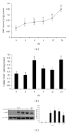
Previous studies from this laboratory demonstrate that dietary leucine protects against high fat diet-induced mitochondrial impairments and stimulates mitochondrial biogenesis and energy partitioning from adipocytes to muscle cells through SIRT1-mediated mechanisms. Moreover, β-hydroxy-β-methyl butyrate (HMB), a metabolite of leucine, has been reported to activate AMPK synergistically with resveratrol in C2C12 myotubes. Therefore, we hypothesize that leucine-induced activation of SIRT1 and AMPK is the central event that links the upregulated mitochondrial biogenesis and fatty acid oxidation in skeletal muscle. Thus, C2C12 myotubes were treated with leucine (0.5 mM), alanine (0.5 mM), valine (0.5 mM), EX527 (SIRT1 inhibitor, 25 μM), and Compound C (AMPK inhibitor, 25 μM) alone or in combination to determine the roles of AMPK and SIRT1 in leucine-modulation of energy metabolism. Leucine significantly increased mitochondrial content, mitochondrial biogenesis-related genes expression, fatty acid oxidation, SIRT1 activity and gene expression, and AMPK phosphorylation in C2C12 myotubes compared to the controls, while EX527 and Compound C markedly attenuated these effects. Furthermore, leucine treatment for 24 hours resulted in time-dependent increases in cellular NAD(+), SIRT1 activity, and p-AMPK level, with SIRT1 activation preceding that of AMPK, indicating that leucine activation of SIRT1, rather than AMPK, is the primary event.
Figures


Similar articles
-
Activation of the AMPK/Sirt1 pathway by a leucine-metformin combination increases insulin sensitivity in skeletal muscle, and stimulates glucose and lipid metabolism and increases life span in Caenorhabditis elegans.Metabolism. 2016 Nov;65(11):1679-1691. doi: 10.1016/j.metabol.2016.06.011. Epub 2016 Jul 9. Metabolism. 2016. PMID: 27456392
-
Leucine regulates slow-twitch muscle fibers expression and mitochondrial function by Sirt1/AMPK signaling in porcine skeletal muscle satellite cells.Anim Sci J. 2019 Feb;90(2):255-263. doi: 10.1111/asj.13146. Epub 2018 Dec 6. Anim Sci J. 2019. PMID: 30523660
-
Synergistic effects of polyphenols and methylxanthines with Leucine on AMPK/Sirtuin-mediated metabolism in muscle cells and adipocytes.PLoS One. 2014 Feb 14;9(2):e89166. doi: 10.1371/journal.pone.0089166. eCollection 2014. PLoS One. 2014. PMID: 24551237 Free PMC article.
-
Synergistic effects of leucine and resveratrol on insulin sensitivity and fat metabolism in adipocytes and mice.Nutr Metab (Lond). 2012 Aug 22;9(1):77. doi: 10.1186/1743-7075-9-77. Nutr Metab (Lond). 2012. PMID: 22913271 Free PMC article.
-
Activation of SIRT1 by L-serine increases fatty acid oxidation and reverses insulin resistance in C2C12 myotubes.Cell Biol Toxicol. 2019 Oct;35(5):457-470. doi: 10.1007/s10565-019-09463-x. Epub 2019 Feb 5. Cell Biol Toxicol. 2019. PMID: 30721374
Cited by
-
Reviewing the Effects of L-Leucine Supplementation in the Regulation of Food Intake, Energy Balance, and Glucose Homeostasis.Nutrients. 2015 May 22;7(5):3914-37. doi: 10.3390/nu7053914. Nutrients. 2015. PMID: 26007339 Free PMC article. Review.
-
Coordinated Modulation of Energy Metabolism and Inflammation by Branched-Chain Amino Acids and Fatty Acids.Front Endocrinol (Lausanne). 2020 Sep 8;11:617. doi: 10.3389/fendo.2020.00617. eCollection 2020. Front Endocrinol (Lausanne). 2020. PMID: 33013697 Free PMC article. Review.
-
Involvement of Mfn2, Bcl2/Bax signaling and mitochondrial viability in the potential protective effect of Royal jelly against mitochondria-mediated ovarian apoptosis by cisplatin in rats.Iran J Basic Med Sci. 2020 Apr;23(4):515-526. doi: 10.22038/ijbms.2020.40401.9563. Iran J Basic Med Sci. 2020. PMID: 32489567 Free PMC article.
-
Uridine alleviates high-carbohydrate diet-induced metabolic syndromes by activating sirt1/AMPK signaling pathway and promoting glycogen synthesis in Nile tilapia (Oreochromis niloticus).Anim Nutr. 2023 May 3;14:56-66. doi: 10.1016/j.aninu.2023.03.010. eCollection 2023 Sep. Anim Nutr. 2023. PMID: 37252330 Free PMC article.
-
Leucine-rich diet induces a shift in tumour metabolism from glycolytic towards oxidative phosphorylation, reducing glucose consumption and metastasis in Walker-256 tumour-bearing rats.Sci Rep. 2019 Oct 29;9(1):15529. doi: 10.1038/s41598-019-52112-w. Sci Rep. 2019. PMID: 31664147 Free PMC article.
References
-
- Schrauwen P, Schrauwen-Hinderling V, Hoeks J, Hesselink MKC. Mitochondrial dysfunction and lipotoxicity. Biochimica et Biophysica Acta. 2010;1801(3):266–271. - PubMed
-
- Howitz KT, Bitterman KJ, Cohen HY, et al. Small molecule activators of sirtuins extend Saccharomyces cerevisiae lifespan. Nature. 2003;425(6954):191–196. - PubMed
LinkOut - more resources
Full Text Sources
Other Literature Sources
Medical

