. 2014 Aug 22;5(9):3140-9.
doi: 10.1364/BOE.5.003140. eCollection 2014 Sep 1.

Preparation of a skin equivalent phantom with interior micron-scale vessel structures for optical imaging experiments

Affiliations

Preparation of a skin equivalent phantom with interior micron-scale vessel structures for optical imaging experiments

Chen Chen et al. Biomed Opt Express. .

Erratum in

Abstract

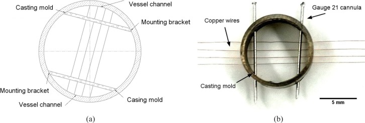
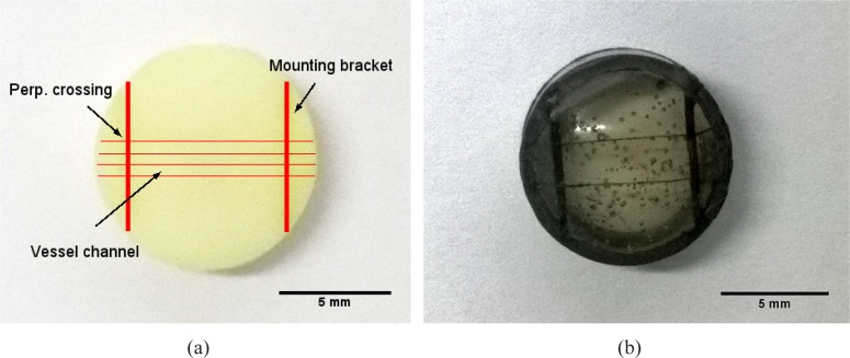
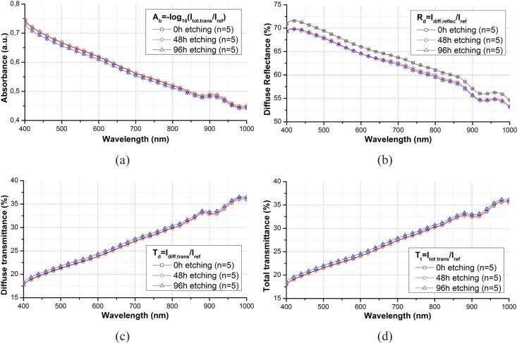
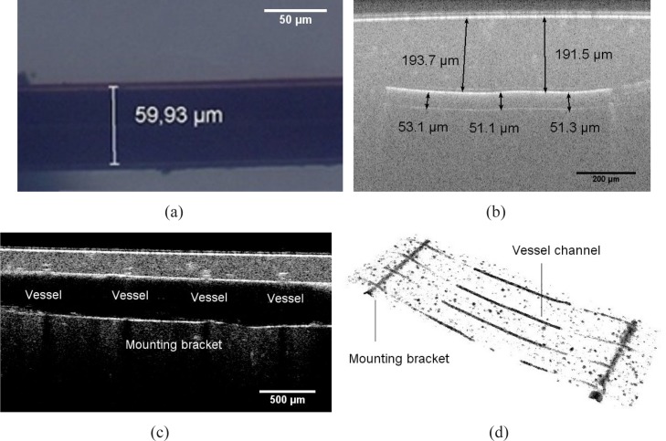
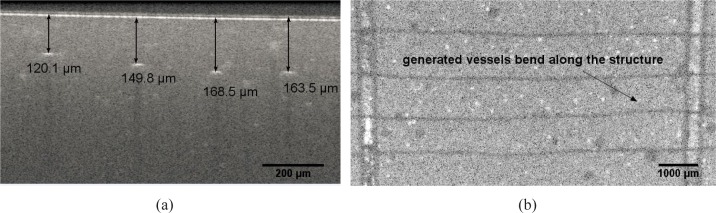
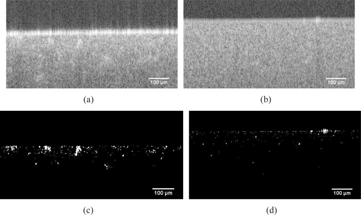
A popular alternative of preparing multilayer or microfluidic chip based phantoms could have helped to simulate the subsurface vascular network, but brought inevitable problems. In this work, we describe the preparation method of a single layer skin equivalent tissue phantom containing interior vessel channels, which mimick the superficial microvascular structure. The fabrication method does not disturb the optical properties of the turbiding matrix material. The diameter of the channels reaches a value of 50 μm. The size, as well as the geometry of the generated vessel structures are investigated by using the SD-OCT system. Our preliminary results confirm that fabrication of such a phantom is achievable and reproducible. Prospectively, this phantom is used to calibrate the optical angiographic imaging approaches.

Keywords: (110.7050) Turbid media; (160.4760) Optical properties; (170.0170) Medical optics and biotechnology; (170.3880) Medical and biological imaging; (350.0350) Other areas of optics.


Figures

Fig. 1
Fig. 1
(a) Schematic of the cylinder casting mold design for regulating 4 rows of vessel channels in the tissue phantom and (b) image of a finished casting mold with 4 rows of copper wires and 21 gauge cannulas inserted through the drilling holes
Fig. 2
Fig. 2
Picture of (a) a finished tissue phantom with 4 rows of vessel channels perpendicularly crossing over 2 mounting brackets and (b) a finished transparent tissue phantom with 2 rows of channels, they are included with india ink to highlight the hollow vessel channels against the matrix material
Fig. 3
Fig. 3
Comparison of (a) absorbance, (b) diffuse reflectance (c) diffuse transmittance and (d) total transmittance spectra through a phantom slab before and after etching in wavelength range from 400 to 1000 nm and their standard derivation.
Fig. 4
Fig. 4
(a) OM graphic of a generated vessel channel dyed with india ink in a transparent tissue phantom (b) OCT B-scan image of the cross section passing along a vessel channel (c) OCT B-scan image of the cross section passing along a mounting bracket across the the vessel channels (d) OCT volume view of the segmentated vessel structures, including vessel channels and mounting brackets
Fig. 5
Fig. 5
(a) OCT image of the cross section across over 4 vessel channel, showing that they are not located on the same latitude of around 160 μm to the surface (b) OCT top view of vessel channels in tissue phantom : vessel channels bend in tangential direction along the structure due to the inner stress induced during solidification
Fig. 6
Fig. 6
Origin OCT B-Scan image of the cross section of (a) a typically inhomogeneous tissue phantom (b) homogenous tissue phantom, subfigures (c) and (d) demonstrate the segmentated scattering clusters corresponding to (a) and (b)

References

    1. Firbank M., Delpy D. T., “A design for a stable and reproducible phantom for use in near infra-red imaging and spectroscopy,” Phys. Med. Biol. 38(6), 847 (1993)10.1088/0031-9155/38/6/015 - DOI - PubMed
    1. Pogue B. W., Patterson M. S., “Review of tissue simulating phantoms for optical spectroscopy, imaging and dosimetry,” J. Biomed. Opt. 11(4), 041102 (2006)10.1117/1.2335429 - DOI - PubMed
    1. Lualdi M., Colombo A., Farina B., Tomatis S., Marchesini R., “A phantom with tissue-like optical properties in the visible and near infrared for use in photomedicine,” Laser. Surg. Med. 39(3), 237–243 (2001)10.1002/lsm.1044 - DOI - PubMed
    1. Roystonand D. D., Poston R. S., Prahl S. A., “Optical properties of scattering and absorbing materials used in the development of optical phantoms at 1064 nm,” J. Biomed. Opt. 1(1), 110–116 (1996)10.1117/12.227698 - DOI - PubMed
    1. Lamouche G., Kennedy B. F., Kennedy K. M., Bisaillon C. E., Curatolo A., Campbell G., Pazos V., Sampson D. D., “Review of tissue simulating phantoms with controllable optical, mechanical and structural properties for use in optical coherence tomography,” Biomed. Opt. Express 3(6), 1381–1398 (2012)10.1364/BOE.3.001381 - DOI - PMC - PubMed