Review
. 2014 Dec 6;11(101):20140459.
doi: 10.1098/rsif.2014.0459.

Liposomes in tissue engineering and regenerative medicine

Review

Liposomes in tissue engineering and regenerative medicine

Nelson Monteiro et al. J R Soc Interface. .

Abstract

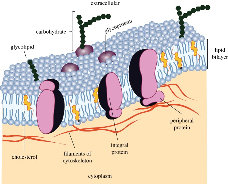
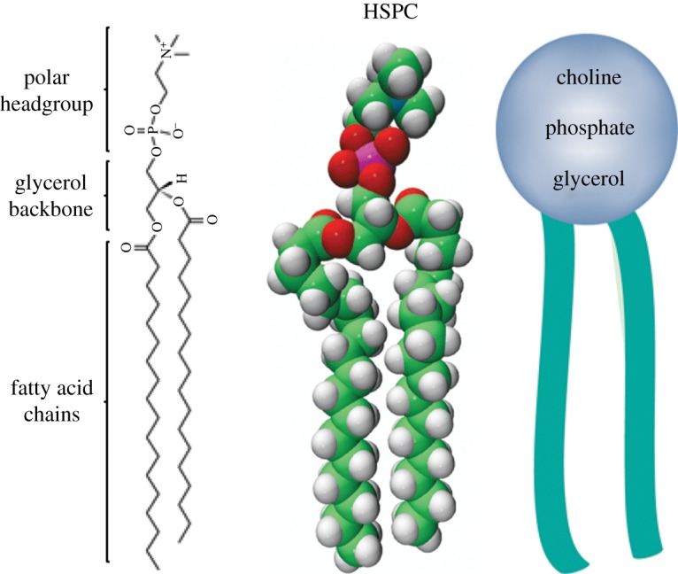
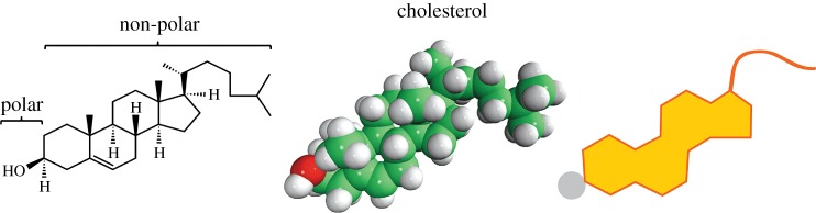
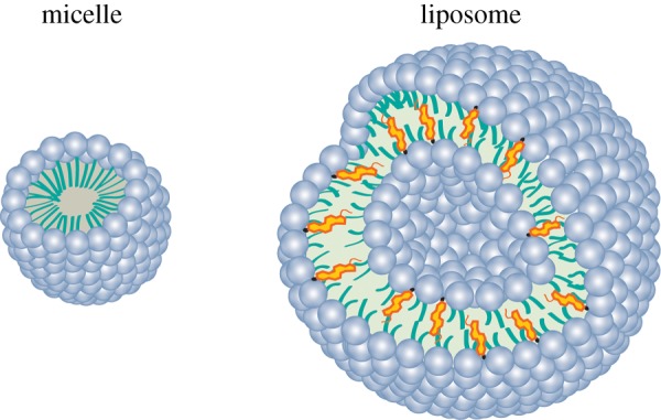
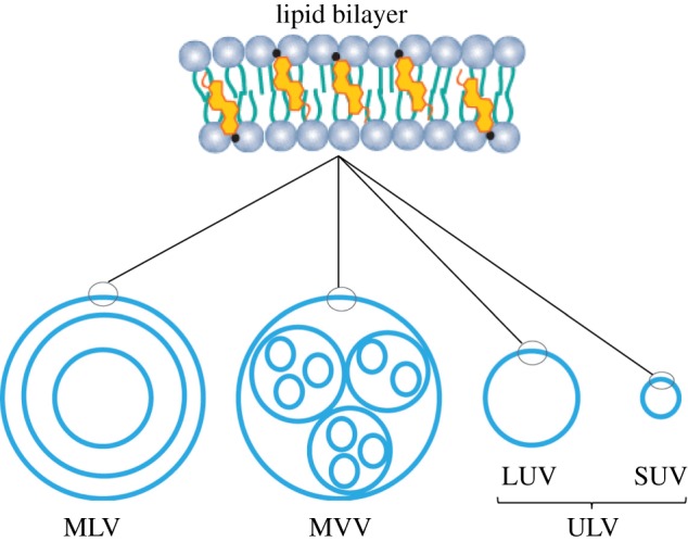
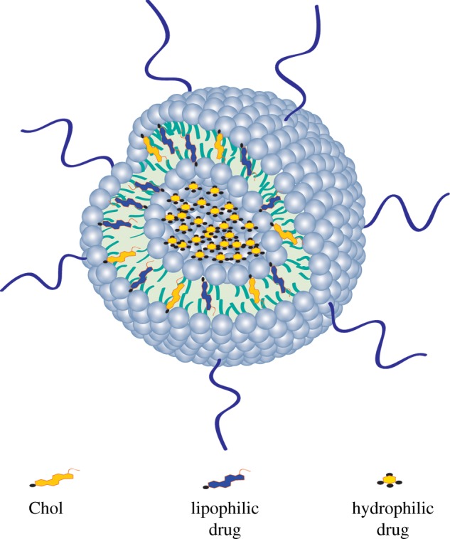
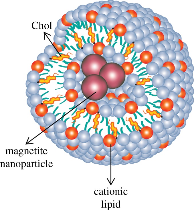
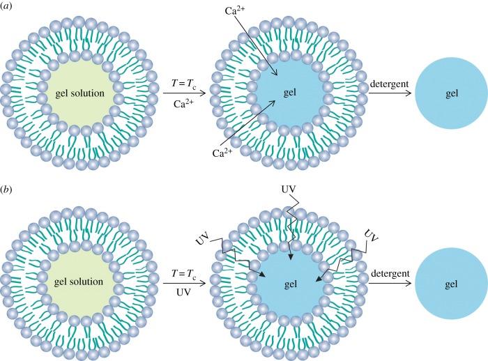
Liposomes are vesicular structures made of lipids that are formed in aqueous solutions. Structurally, they resemble the lipid membrane of living cells. Therefore, they have been widely investigated, since the 1960s, as models to study the cell membrane, and as carriers for protection and/or delivery of bioactive agents. They have been used in different areas of research including vaccines, imaging, applications in cosmetics and tissue engineering. Tissue engineering is defined as a strategy for promoting the regeneration of tissues for the human body. This strategy may involve the coordinated application of defined cell types with structured biomaterial scaffolds to produce living structures. To create a new tissue, based on this strategy, a controlled stimulation of cultured cells is needed, through a systematic combination of bioactive agents and mechanical signals. In this review, we highlight the potential role of liposomes as a platform for the sustained and local delivery of bioactive agents for tissue engineering and regenerative medicine approaches.


Figures

Figure 1.
Figure 1.
A cell membrane is a fluid with various proteins attached to the lipid bilayer. Adapted from [5]. (Online version in colour.)
Figure 2.
Figure 2.
Chemical, three-dimensional and schematic structure of L-α-phosphatidylcholine, hydrogenated (soy) (HSPC) composed of fatty acid chains, glycerol backbone and the headgroup (choline). Adapted from [11]. (Online version in colour.)
Figure 3.
Figure 3.
Chemical, three-dimensional and schematic structure of cholesterol (Chol). Adapted from [14]. (Online version in colour.)
Figure 4.
Figure 4.
Alternative lipid-based particles: micelle and a liposome. (Online version in colour.)
Figure 5.
Figure 5.
Selective permeability of lipid bilayers. Adapted from [14]. (Online version in colour.)
Figure 6.
Figure 6.
Unsaturated and saturated hydrocarbons. A double bond in a hydrocarbon chain creates spaces in the double layer. Adapted from [14]. (Online version in colour.)
Figure 7.
Figure 7.
Effect of temperature and Chol on phospholipid bilayer permeability. Chol eliminates the transition phase of the lipid bilayer. The Chol increases the lipid bilayer permeability in the gel phase and decreases it in the fluid phase. Adapted from [15,35]. (Online version in colour.)
Figure 8.
Figure 8.
Cross section of a liposome: (a) conventional liposome; (b) SSL; (c) ligand-targeted liposome; (d) fluorescent liposome and charged liposomes. (Online version in colour.)
Figure 9.
Figure 9.
Lipid bilayer structure and types of liposomes: MLVs, MVVs, ULVs. Additionally, ULVs can be sub-classified as LUVs and SUVs. Adapted from [18]. (Online version in colour.)
Figure 10.
Figure 10.
Representation of hydrophilic and lipophilic drug encapsulation into the liposome. Hydrophilic drug is encapsulated in the inner core of the liposome. Lipophilic drug is encapsulated into the lipid bilayer. Chol and the lipophilic drug compete for the same space in the lipid bilayer. (Online version in colour.)
Figure 11.
Figure 11.
Magnetite cationic liposome. (Online version in colour.)
Figure 12.
Figure 12.
Strategies to produce nanogels using liposomes as a template. (a) Ca2+ calcium alginate gelation and (b) UV-photopolymerization. (Online version in colour.)

References

    1. Fahy E, et al. 2009. Update of the LIPID MAPS comprehensive classification system for lipids. J. Lipid Res. 50, S9–S14. (10.1194/jlr.R800095-JLR200) - DOI - PMC - PubMed
    1. LIPID-MAPS. 2014. Lipidomics gateway See http://www.lipidmaps.org.
    1. Vemuri S, Rhodes CT. 1995. Preparation and characterization of liposomes as therapeutic delivery systems: a review. Pharm. Acta Helv. 70, 95–111. (10.1016/0031-6865(95)00010-7) - DOI - PubMed
    1. Subramaniam S, Fahy E, Gupta S, Sud M, Byrnes RW, Cotter D, Dinasarapu AR, Maurya MR. 2011. Bioinformatics and systems biology of the lipidome. Chem. Rev. 111, 6452–6490. (10.1021/cr200295k) - DOI - PMC - PubMed
    1. Mader S. 2010. Inquiry into life, 13, ilustrada edn New York, NY: McGraw-Hill Education.

Publication types

MeSH terms