

Differentiation of retinal ganglion cells and photoreceptor precursors from mouse induced pluripotent stem cells carrying an Atoh7/Math5 lineage reporter
- PMID: 25401462
- PMCID: PMC4234374
- DOI: 10.1371/journal.pone.0112175
Differentiation of retinal ganglion cells and photoreceptor precursors from mouse induced pluripotent stem cells carrying an Atoh7/Math5 lineage reporter
Abstract
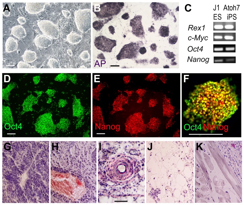
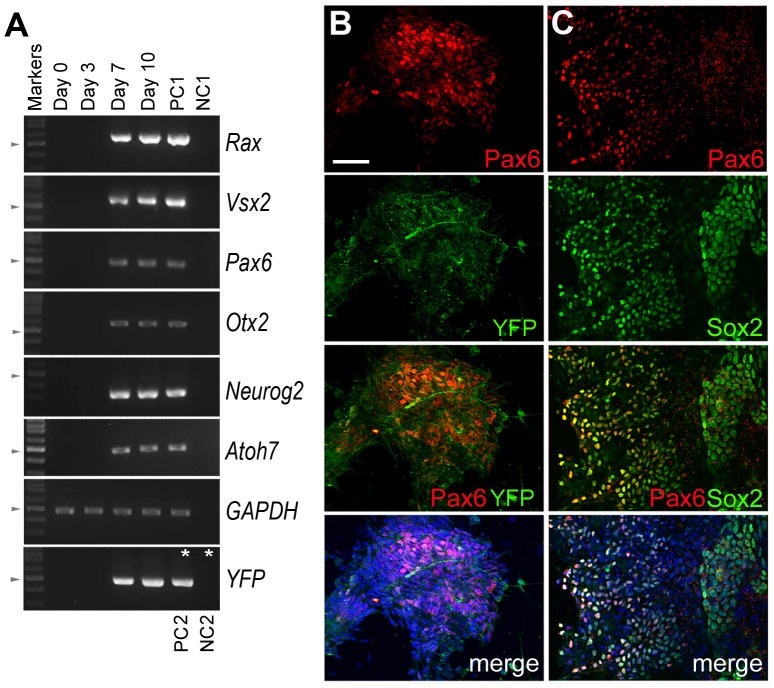
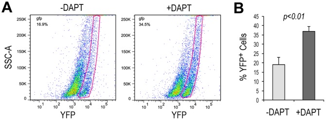
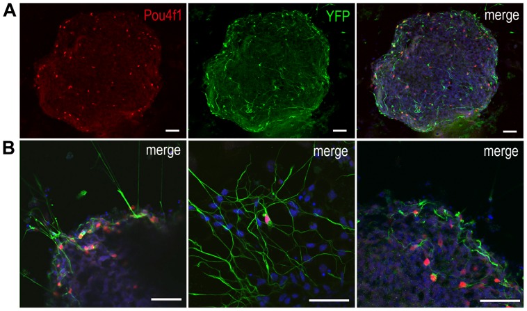
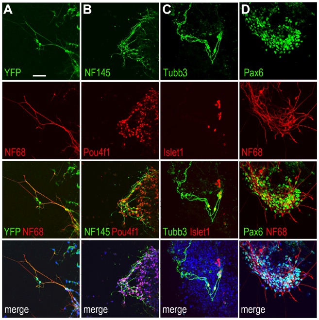
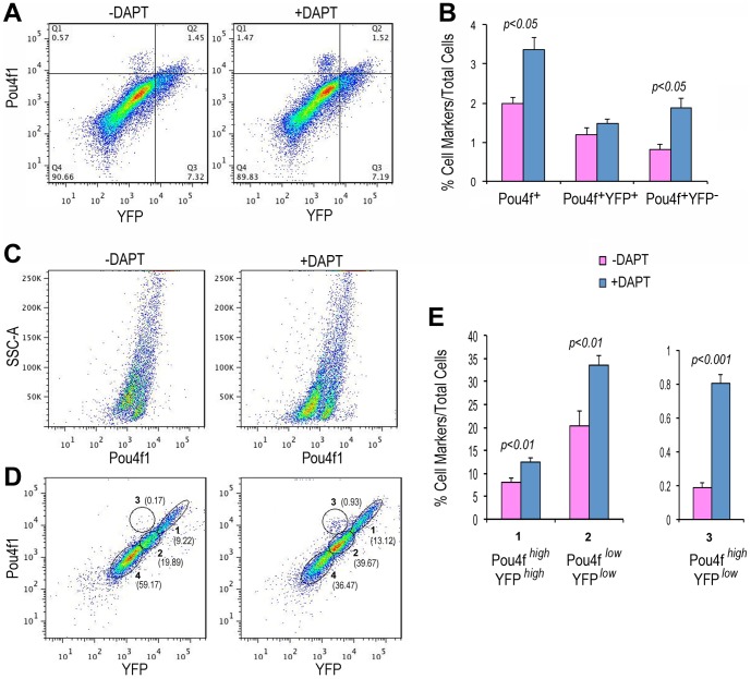
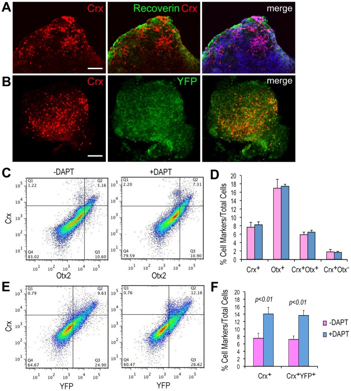
The neural retina is a critical component of the visual system, which provides the majority of sensory input in humans. Various retinal degenerative diseases can result in the permanent loss of retinal neurons, especially the light-sensing photoreceptors and the centrally projecting retinal ganglion cells (RGCs). The replenishment of lost RGCs and the repair of optic nerve damage are particularly challenging, as both RGC specification and their subsequent axonal growth and projection involve complex and precise regulation. To explore the developmental potential of pluripotent stem cell-derived neural progenitors, we have established mouse iPS cells that allow cell lineage tracing of progenitors that have expressed Atoh7/Math5, a bHLH transcription factor required for RGC production. These Atoh7 lineage reporter iPS cells encode Cre to replace one copy of the endogenous Atoh7 gene and a Cre-dependent YFP reporter in the ROSA locus. In addition, they express pluripotent markers and are capable of generating teratomas in vivo. Under anterior neural induction and neurogenic conditions in vitro, the Atoh7-Cre/ROSA-YFP iPS cells differentiate into neurons that co-express various RGC markers and YFP, indicating that these neurons are derived from Atoh7-expressing progenitors. Consistent with previous in vivo cell lineage studies, the Atoh7-Cre/ROSA-YFP iPS cells also give rise to a subset of Crx-positive photoreceptor precursors. Furthermore, inhibition of Notch signaling in the iPSC cultures results in a significant increase of YFP-positive RGCs and photoreceptor precursors. Together, these results show that Atoh7-Cre/ROSA-YFP iPS cells can be used to monitor the development and survival of RGCs and photoreceptors from pluripotent stem cells.
Conflict of interest statement
Figures
Similar articles
-
Pushing the envelope of retinal ganglion cell genesis: context dependent function of Math5 (Atoh7).Dev Biol. 2012 Aug 15;368(2):214-30. doi: 10.1016/j.ydbio.2012.05.005. Epub 2012 May 15. Dev Biol. 2012. PMID: 22609278 Free PMC article.
-
Math5 defines the ganglion cell competence state in a subpopulation of retinal progenitor cells exiting the cell cycle.Dev Biol. 2012 May 15;365(2):395-413. doi: 10.1016/j.ydbio.2012.03.006. Epub 2012 Mar 15. Dev Biol. 2012. PMID: 22445509 Free PMC article.
-
Reprogramming amacrine and photoreceptor progenitors into retinal ganglion cells by replacing Neurod1 with Atoh7.Development. 2013 Feb 1;140(3):541-51. doi: 10.1242/dev.085886. Development. 2013. PMID: 23293286 Free PMC article.
-
Genetic control of retinal ganglion cell genesis.Cell Mol Life Sci. 2021 May;78(9):4417-4433. doi: 10.1007/s00018-021-03814-w. Epub 2021 Mar 29. Cell Mol Life Sci. 2021. PMID: 33782712 Free PMC article. Review.
-
Making neurons, made easy: The use of Neurogenin-2 in neuronal differentiation.Stem Cell Reports. 2022 Jan 11;17(1):14-34. doi: 10.1016/j.stemcr.2021.11.015. Epub 2021 Dec 30. Stem Cell Reports. 2022. PMID: 34971564 Free PMC article. Review.
Cited by
-
Directly induced human retinal ganglion cells mimic fetal RGCs and are neuroprotective after transplantation in vivo.Stem Cell Reports. 2022 Dec 13;17(12):2690-2703. doi: 10.1016/j.stemcr.2022.10.011. Epub 2022 Nov 10. Stem Cell Reports. 2022. PMID: 36368332 Free PMC article.
-
Differentiation of retinal ganglion cells from induced pluripotent stem cells: a review.Int J Ophthalmol. 2019 Jan 18;12(1):152-160. doi: 10.18240/ijo.2019.01.22. eCollection 2019. Int J Ophthalmol. 2019. PMID: 30662854 Free PMC article. Review.
-
Differentiation of Human Embryonic/Induced-Pluripotent Stem Cells to Retinal Ganglion Cells.Methods Mol Biol. 2023;2708:41-48. doi: 10.1007/978-1-0716-3409-7_5. Methods Mol Biol. 2023. PMID: 37558958
-
Blind But Alive - Congenital Loss of atoh7 Disrupts the Visual System of Adult Zebrafish.Invest Ophthalmol Vis Sci. 2024 Nov 4;65(13):42. doi: 10.1167/iovs.65.13.42. Invest Ophthalmol Vis Sci. 2024. PMID: 39565303 Free PMC article.
-
Inherited eye-related disorders due to mitochondrial dysfunction.Hum Mol Genet. 2017 Aug 1;26(R1):R12-R20. doi: 10.1093/hmg/ddx182. Hum Mol Genet. 2017. PMID: 28481993 Free PMC article. Review.
References
-
- Turner DL, Cepko CL (1987) A common progenitor for neurons and glia persists in rat retina late in development. Nature 328: 131–136. - PubMed
-
- Holt CE, Bertsch TW, Ellis HM, Harris WA (1988) Cellular determination in the Xenopus retina is independent of lineage and birth date. Neuron 1: 15–26. - PubMed
-
- Wetts R, Fraser SE (1988) Multipotent precursors can give rise to all major cell types of the frog retina. Science 239: 1142–1145. - PubMed
-
- Hyde DR, Reh TA (2014) The past, present, and future of retinal regeneration. Exp Eye Res. - PubMed
Publication types
MeSH terms
Substances
Grants and funding
LinkOut - more resources
Full Text Sources
Other Literature Sources
Molecular Biology Databases
