Clinical Trial
. 2014 Dec;124(12):5323-36.
doi: 10.1172/JCI76629. Epub 2014 Nov 17.

Human glial chimeric mice reveal astrocytic dependence of JC virus infection

Clinical Trial

Human glial chimeric mice reveal astrocytic dependence of JC virus infection

Yoichi Kondo et al. J Clin Invest. 2014 Dec.

Abstract

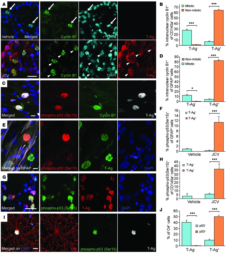
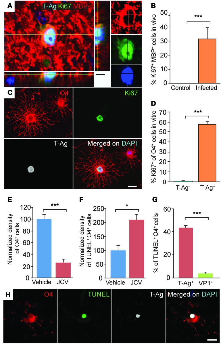
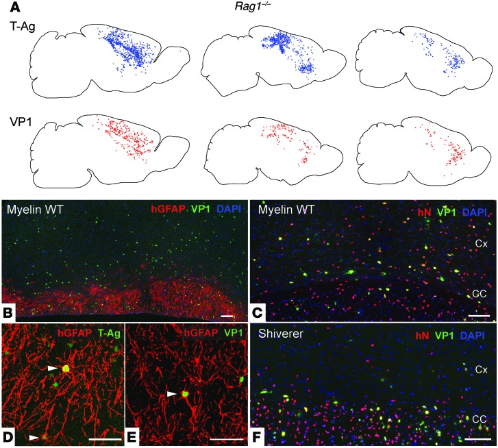
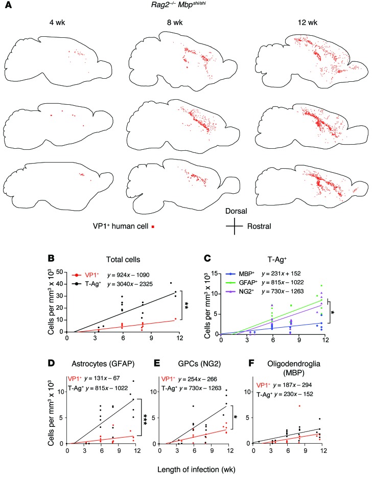
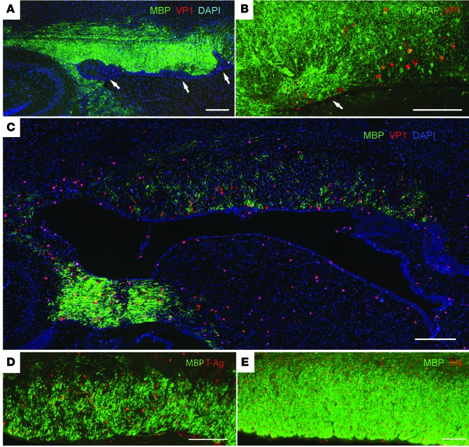
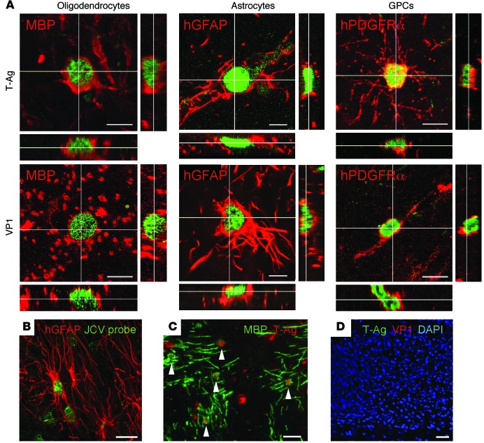
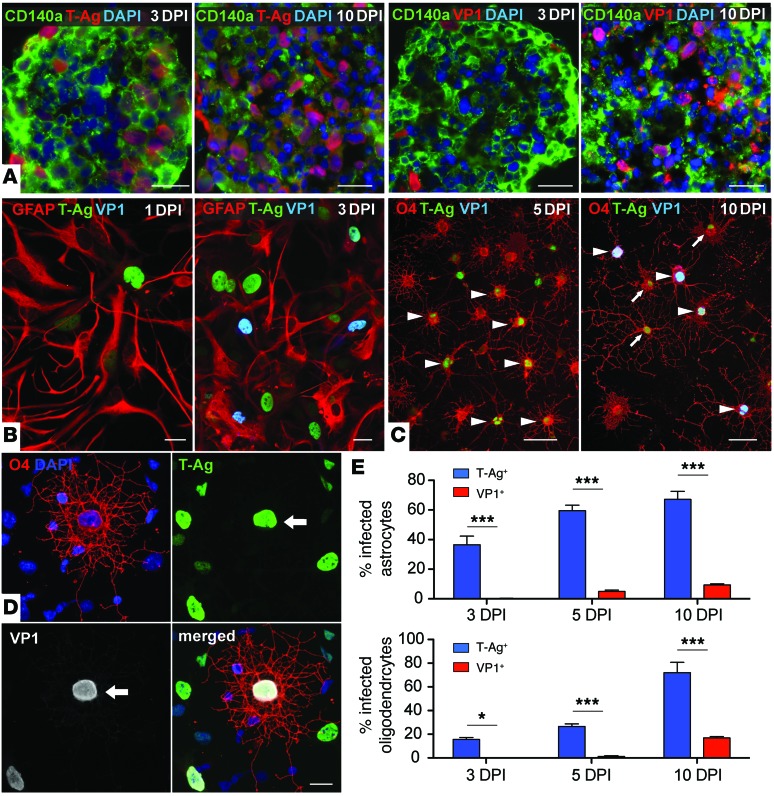
Progressive multifocal leukoencephalopathy (PML) is a demyelinating disease triggered by infection with the human gliotropic JC virus (JCV). Due to the human-selective nature of the virus, there are no animal models available to investigate JCV pathogenesis. To address this issue, we developed mice with humanized white matter by engrafting human glial progenitor cells (GPCs) into neonatal immunodeficient and myelin-deficient mice. Intracerebral delivery of JCV resulted in infection and subsequent demyelination of these chimeric mice. Human GPCs and astrocytes were infected more readily than oligodendrocytes, and viral replication was noted primarily in human astrocytes and GPCs rather than oligodendrocytes, which instead expressed early viral T antigens and exhibited apoptotic death. Engraftment of human GPCs in normally myelinated and immunodeficient mice resulted in humanized white matter that was chimeric for human astrocytes and GPCs. JCV effectively propagated in these mice, which indicates that astroglial infection is sufficient for JCV spread. Sequencing revealed progressive mutation of the JCV capsid protein VP1 after infection, suggesting that PML may evolve with active infection. These results indicate that the principal CNS targets for JCV infection are astrocytes and GPCs and that infection is associated with progressive mutation, while demyelination is a secondary occurrence, following T antigen-triggered oligodendroglial apoptosis. More broadly, this study provides a model by which to further assess the biology and treatment of human-specific gliotropic viruses.


Figures

Figure 7
Figure 7. Cell cycle arrest at G2/M in infected glia.
T-Ag+ glia expressed the mitosis-associated nuclear protein cyclin B1, as well as the DNA damage and cell cycle arrest–associated phospho-p53(Ser15), regardless of their mitotic stage. (A and B) Vehicle-treated and uninfected (T-Ag) GPCs expressed intranuclear cyclin B1 only when mitotic and in M phase (the latter as assessed by DAPI; arrows), whereas the nuclei of JCV-infected (T-Ag+) GPCs admitted cyclin B1 in a temporally promiscuous fashion (arrowheads). (C) Nuclear phospho-p53(Ser15) and cyclin B1 were coexpressed by mitotically arrested infected astrocytes, but not by uninfected astrocytes. (D) Like control GPCs, uninfected astrocytes (T-AgGFAP+) expressed intranuclear cyclin B1 only in M phase, whereas JCV-infected astrocytes expressed nuclear cyclin B1 even when not dividing. (E and F) JCV-infected astrocytes (T-Ag+GFAP+) coexpressed phospho-p53(Ser15), associated with G/2M arrest, whereas neither vehicle-treated nor T-Ag astroglia in infected cultures did so to any significant degree. (G and H) Similarly, JCV-infected GPCs (T-Ag+CD140a+) coexpressed phospho-p53(Ser15), as well as did infected oligodendrocytes (T-Ag+O4+) (I and J); for both oligodendrocytes and their progenitors, T-Ag+ cells were significantly more likely to express phospho-p53(Ser15). All cultures were assessed at 10 DPI with type 2A JCV (Mad-1 NCCR). Scale bars: 20 μm. *P < 0.05; ***P < 0.001.
Figure 6
Figure 6. JCV-infection leads to cell cycle entry-associated with oligodendrocytic death.
(A and B) Mad-1 JCV-infected MBP+ oligodendrocytes were frequently noted to coexpress Ki67, a marker of mitotic entry, suggesting the aberrant entry of these typically postmitotic cells into cell cycle. (A) Representative T-Ag+Ki67+MBP+ oligodendrocyte in the corpus callosum of a human glial chimeric Rag2–/– Mbpshi/shi mouse 12 weeks after infection. (B) Whereas roughly one-third of all infected MBP+ oligodendroglia were Ki67+ at this time point, no Ki67+MBP+ oligodendrocytes were noted in uninfected controls. (C and D) T-Ag+Ki67+O4+ oligodendrocytes were common in vitro (C), and most T-Ag+ oligodendroglia were Ki67+, while few if any uninfected (T-Ag) O4+ oligodendroglia were Ki67+ (D). In culture, JCV infection significantly reduced the number of O4+ oligodendrocytes at 10 DPI (E) by inducing TUNEL-defined apoptosis (F). At 10 DPI, of infected TUNEL+O4+ oligodendrocytes, 43.5% ± 2.1% were T-Ag+, whereas only 4.6% ± 0.3% were VP1+ (G), indicative of the failure of most infected oligodendroglia to progress to VP1-defined viral replication before dying. (H) TUNEL expression by T-Ag+ pyknotic O4+ oligodendrocytes was typical. *P < 0.05, ***P < 0.001, paired 2-tailed Student’s t test. (B) n = 3 per group. (DG) n = 4 runs, triplicate wells; >2,000 scored cells/group. Scale bars: 10 μm (A); 20 μm (C and H).
Figure 5
Figure 5. Astrocytes and GPCs are sufficient to support viral replication and spread in vivo.
(A) JCV introduced into myelin WT Rag1–/– mice (colonized with human GPCs and astrocytes, but not oligodendrocytes) yielded viral propagation and geographic spread as rapid and extensive as that in human glial chimeric Rag2–/– Mbpshi/shi mice (in which human oligodendroglia were densely represented). Shown are distributions of T-Ag+ and VP1+ cells mapped in 14-μm sagittal sections of 3 different Rag1–/– mice injected with JCV as adults, 12 weeks previously. Infected human cells were widely distributed, despite the absence of human oligodendroglia. (B) Sagittal section along the callosal length of a chimeric myelin WT Rag1–/– mouse 12 weeks after infection, showing widespread infection and VP1 expression by GFAP+ subcortical human astrocytes and GFAP cortical human astrocytes and GPCs. (D and E) Higher-magnification views showing the predominance of infected cells (arrowheads) in Rag1–/– cortical grey, including both T-Ag+ (D) and VP1+ (E) glia, manifesting the typical hypertrophic nuclei of cells that have undergone viral replication. (F) Conversely, VP1+ glia in the corpus callosum of a human glial chimeric Rag2–/– Mbpshi/shi mouse 12 weeks after type 1A (Mad-1) JCV infection showed the predominant white matter spread of virus in these mice, which manifested both oligodendrocytic and astrocytic infection (compare with the Rag1–/– section in C). Notably, since JCV can only infect human cells, the potential volume of infection in these mice was limited by the geographic dispersal of the resident human oligodendrocyte progenitor cells. Since these mice were given only forebrain injections so as to produce forebrain-only human glial chimeras, all JCV infection in these mice was limited to the forebrain. Cx, cerebral cortex; CC, corpus callosum. Scale bars: 100 μm (B, C, and F); 50 μm (D and E).
Figure 4
Figure 4. Viral propagation exhibits cell type–selective spread.
JCV spread in vivo was tracked by immunostaining human glial chimeric Rag2–/– Mbpshi/shi brains for both T-Ag and VP1, each as a function of time after infection. In these chimeric Rag2–/– Mbpshi/shi mice, a large proportion of oligodendrocytes, as well as GPCs and astrocytes, were human. (A) Sagittal sections of 3 different infected chimeras at each of 3 time points; individual VP1+ cells are dot-mapped. VP1+ human cells became progressively more widespread with time, with JCV infection progressing from the site of viral injection to include much of the forebrain white matter by 12 weeks postinfection, with marked cortical spread by that point as well. (B) T-Ag+ and VP1+ cells, representing all JCV-infected cells and those in which viral replication has occurred, respectively, both accumulated as a function of time. (C) The number of T-Ag+ astrocytes (GFAP+) and GPCs (NG2+) was significantly higher than that of T-Ag+ oligodendroglia (MBP+) at all time points examined (P < 0.01, repeated-measures 1-way ANOVA). Furthermore, the in vivo rates of accumulation of T-Ag+ astrocytes and GPCs (reflecting the rate of infection among each cell type, estimated by regression line slope) were each significantly higher than that of oligodendrocytes (P < 0.01, linear regression). (DF) Despite their marked differences in T-Ag–defined infectivity and spread, astrocytes and oligodendrocytes did not differ in their rates of accumulation of VP1+ infectants, likely reflecting the rapid lytic loss of cells at that stage. *P < 0.05; **P < 0.01; ***P < 0.001.
Figure 3
Figure 3. JCV infection of human glial chimeras triggers both focal and diffuse demyelination.
(A and B) At 4 weeks after viral infection, focal regions of demyelination (A, arrows) and infection-associated astrogliosis (B, arrow) were noted in the forebrain white matter of infected mice, typically in discrete foci abutting the callosal wall of the lateral ventricle. hGFAP, human GFAP. (C and D) By 11 weeks after infection, diffuse hypomyelination of the callosa and capsules of infected chimeric mice was noted. (E) Uninfected human chimeric controls by 20 weeks after transplantation exhibited dense GPC-derived myelination, as did JCV-injected non-xenografted murine controls (not shown). hN, human nuclear antigen. Scale bars: 200 μm (A and C); 100 μm (B, D, and E).
Figure 2
Figure 2. JCV replicates more rapidly and efficiently in astroglia than in oligodendrocytes in vivo.
JCV induced the expression of the major early and late viral gene products (T-Ag and VP1 protein, respectively) throughout the corpus callosum of human glial chimeric Rag2–/– Mbpshi/shi mice. (A) Confocal images of infected oligodendrocytes, astrocytes, and GPCs in Rag2–/– Mbpshi/shi mice neonatally engrafted with human GPCs and infected with type 1A (Mad-1) JCV for 12 weeks. (B) By 12 weeks, infected astrocytes were highly abundant and largely magnocellular, with overtly enlarged nuclei and bizarrely fibrotic processes. (C) In contrast, substantial human oligodendrocytic loss was evident by 12 weeks after infection, and most of the infected remainder expressed T-Ag (arrowheads), as exemplified by the T-Ag+MBP+ oligodendroglia shown; only human oligodendroglia expressed MBP in Rag2–/– Mbpshi/shi brain. (D) Infection was restricted to human cells; in this example, unengrafted mouse corpus callosum manifested no evidence of infection 12 weeks after JCV injection. Scale bars: 20 μm (A); 50 μm (B and C); 100 μm (D).
Figure 1
Figure 1. Human astroglia are most efficiently infected by JCV in vitro.
Human GPCs and astrocytes were readily infected by JCV in vitro, with robust expression within days of both the early viral large T-Ag and the later VP1 capsid protein. (A) Both T-Ag and VP1 were expressed by CD140a+ GPCs grown in suspension culture, and both were more abundantly expressed at 10 DPI than at 3 DPI. (B) In CD44-sorted GFAP+ astrocytes, T-Ag was expressed as early as 1 DPI, whereas VP1 was first expressed at 3 DPI. (C) In contrast, O4+ oligodendrocyte infection in vitro was both delayed and of relatively low efficiency, showing weak T-Ag expression without VP1 at 5 DPI (arrowheads), with VP1+ oligodendroglia (arrows) appearing only at 10 DPI. (D) Representative images of a JCV-infected T-Ag+VP1+ oligodendrocyte at 10 DPI; nuclear hypertrophy (arrows) was apparent. (E) At 3 and 5 DPI, T-Ag+ oligodendroglial infection was of significantly lower efficiency than that of astrocytes, while oligodendrocytic VP1+ JCV replication was even less frequent. By 10 DPI, VP1+ oligodendroglia as well as astrocytes began to accumulate. Data are presented as percentage of cells of each phenotype at 3, 5, and 10 DPI. Scale bars: 20 μm. *P < 0.05; ***P < 0.01.

Comment in

References

    1. Berger JR. Progressive multifocal leukoencephalopathy. Current Neurol Neurosci Reports. 2007;7(6):461–469. doi: 10.1007/s11910-007-0072-9. - DOI - PubMed
    1. Major EO. Progressive multifocal leukoencephalopathy in patients on immunomodulatory therapies. Annu Rev Med. 2010;61:35–47. doi: 10.1146/annurev.med.080708.082655. - DOI - PubMed
    1. Tan CS, et al. Detection of JC virus-specific immune responses in a novel humanized mouse model. PloS One. 2013;8(5):e64313. doi: 10.1371/journal.pone.0064313. - DOI - PMC - PubMed
    1. Matoba T, et al. An siRNA against JC virus (JCV) agnoprotein inhibits JCV infection in JCV-producing cells inoculated in nude mice. Neuropathology. 2008;28(3):286–294. - PubMed
    1. Windrem MS, et al. Fetal and adult human oligodendrocyte progenitor cell isolates myelinate the congenitally dysmyelinated brain. Nat Med. 2004;10(1):93–97. doi: 10.1038/nm974. - DOI - PubMed

Publication types

MeSH terms